

Search Count: 24
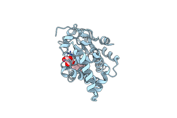 |
Crystal Structure Of The Complex Of The Cellular Retinal Binding Protein With 9-Cis-Retinal
Organism: Homo sapiens
Method: X-RAY DIFFRACTION Resolution:3.40 Å Release Date: 2014-01-08 Classification: ISOMERASE Ligands: RET, TLA |
Organism: Homo sapiens
Method: X-RAY DIFFRACTION
Release Date: 2014-01-08
Ligands: RET, TLA
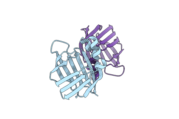 |
Crystal Structure Of The Apo R132K:L121E Mutant Of Cellular Retinoic Acid-Binding Protein Ii At 1.68 Angstrom Resolution
Organism: Homo sapiens
Method: X-RAY DIFFRACTION Resolution:1.68 Å Release Date: 2009-07-28 Classification: lipid binding, retinal binding |
Organism: Homo sapiens
Method: X-RAY DIFFRACTION
Release Date: 2009-07-28
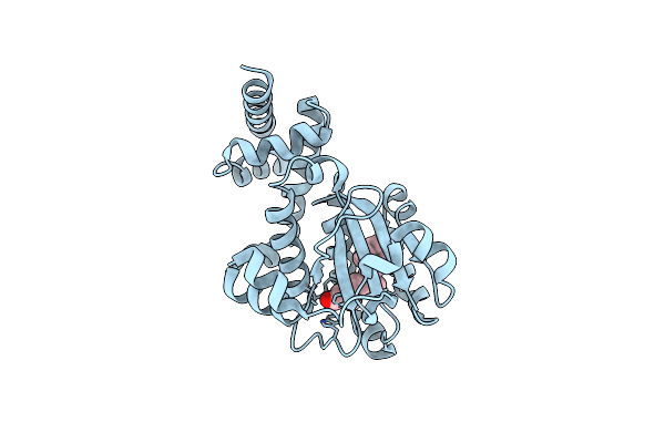 |
Crystal Structure Of The Complex Of The Cellular Retinal Binding Protein Mutant R234W With 9-Cis-Retinal
Organism: Homo sapiens
Method: X-RAY DIFFRACTION Resolution:1.90 Å Release Date: 2014-01-08 Classification: ISOMERASE Ligands: RET |
Organism: Homo sapiens
Method: X-RAY DIFFRACTION
Release Date: 2014-01-08
Ligands: RET
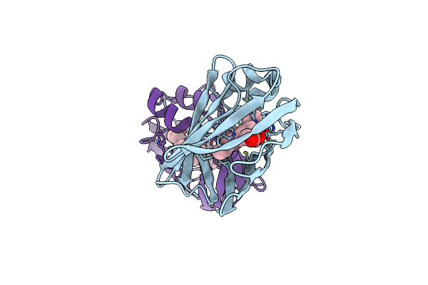 |
Q108K:K40L:T51V:T53C:R58W:Y19W:Q38L:Q4A:T29L Mutant Of Hcrbpii Bound To Fr1V Fluorophore In The Dark State At Ph 4
Organism: Homo sapiens
Method: X-RAY DIFFRACTION Resolution:1.20 Å Release Date: 2025-02-12 Classification: RETINOL BINDING PROTEIN Ligands: ZFJ, ACY |
Organism: Homo sapiens
Method: X-RAY DIFFRACTION
Release Date: 2025-02-12
Ligands: ZFJ, ACY
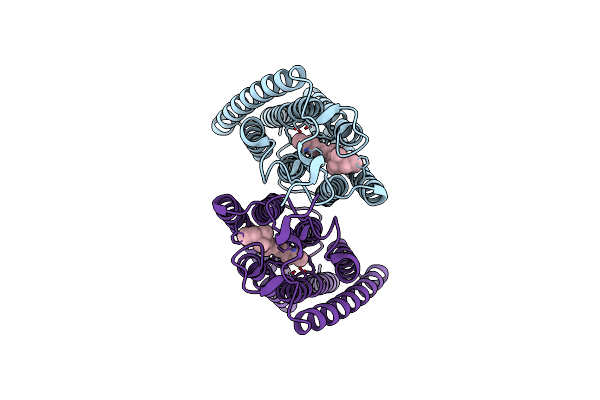 |
Exochr2 Channelrhodopsin
Organism: Chlamydomonas reinhardtii
Method: ELECTRON MICROSCOPY Release Date: 2024-09-04 Classification: MEMBRANE PROTEIN Ligands: RET |
Organism: Chlamydomonas reinhardtii
Method: ELECTRON MICROSCOPY
Release Date: 2024-09-04
Ligands: RET
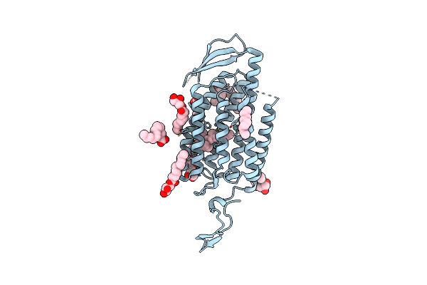 |
C1C2 Channelrhodopsin - Smx Dark Structure
Organism: Chlamydomonas reinhardtii
Method: X-RAY DIFFRACTION Resolution:2.59 Å Release Date: 2025-01-29 Classification: MEMBRANE PROTEIN Ligands: OLC, RET |
Organism: Chlamydomonas reinhardtii
Method: X-RAY DIFFRACTION
Release Date: 2025-01-29
Ligands: OLC, RET
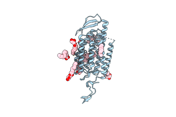 |
C1C2 Channelrhodopsin - Smx Light Activated Structure
Organism: Chlamydomonas reinhardtii
Method: X-RAY DIFFRACTION Resolution:2.70 Å Release Date: 2025-01-29 Classification: MEMBRANE PROTEIN Ligands: OLC, RET |
Organism: Chlamydomonas reinhardtii
Method: X-RAY DIFFRACTION
Release Date: 2025-01-29
Ligands: OLC, RET
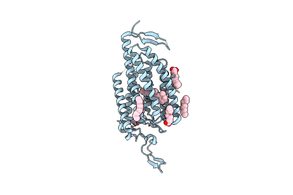 |
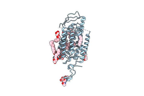
Crystal Structure Of Blue-Shifted Channelrhodopsin Mutant (T198G/G202A)
Organism: Chlamydomonas reinhardtii
Method: X-RAY DIFFRACTION Resolution:2.50 Å Release Date: 2015-05-27 Classification: MEMBRANE PROTEIN Ligands: RET, OLA, ZN |
Organism: Chlamydomonas reinhardtii
Method: X-RAY DIFFRACTION
Release Date: 2015-05-27
Ligands: RET, OLA, ZN
 |
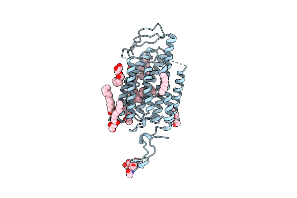
Time-Resolved Serial Femtosecond Crystallography Reveals Early Structural Changes In Channelrhodopsin: 4 Ms Structure
Organism: Chlamydomonas reinhardtii
Method: X-RAY DIFFRACTION Resolution:2.50 Å Release Date: 2021-04-07 Classification: MEMBRANE PROTEIN Ligands: RET, OLC |
Organism: Chlamydomonas reinhardtii
Method: X-RAY DIFFRACTION
Release Date: 2021-04-07
Ligands: RET, OLC
 |
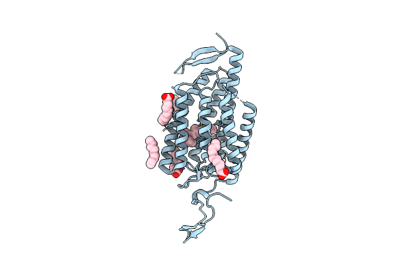
Time-Resolved Serial Femtosecond Crystallography Reveals Early Structural Changes In Channelrhodopsin: 250 Microsecond Structure
Organism: Chlamydomonas reinhardtii
Method: X-RAY DIFFRACTION Resolution:2.50 Å Release Date: 2021-04-07 Classification: MEMBRANE PROTEIN Ligands: RET, OLC |
Organism: Chlamydomonas reinhardtii
Method: X-RAY DIFFRACTION
Release Date: 2021-04-07
Ligands: RET, OLC
 |
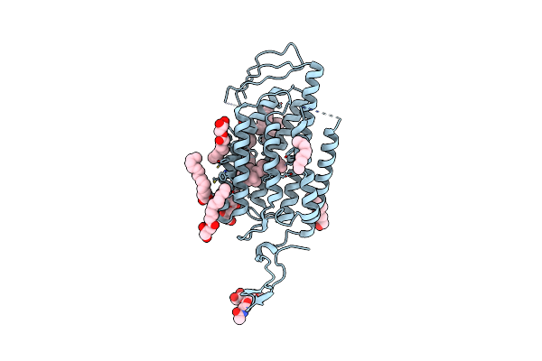
Crystal Structure Of The Closed State Of Channelrhodopsin
Organism: Chlamydomonas reinhardtii
Method: X-RAY DIFFRACTION Resolution:2.30 Å Release Date: 2012-01-25 Classification: MEMBRANE PROTEIN Ligands: RET, OLA |
Organism: Chlamydomonas reinhardtii
Method: X-RAY DIFFRACTION
Release Date: 2012-01-25
Ligands: RET, OLA
 |
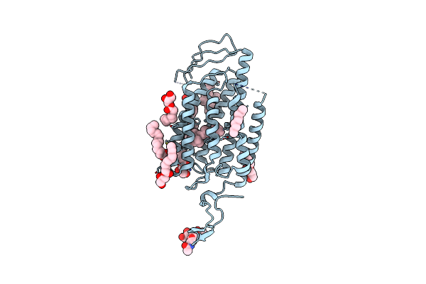
Time-Resolved Serial Femtosecond Crystallography Reveals Early Structural Changes In Channelrhodopsin: 1 Microsecond Structure
Organism: Chlamydomonas reinhardtii
Method: X-RAY DIFFRACTION Resolution:2.50 Å Release Date: 2021-04-07 Classification: MEMBRANE PROTEIN Ligands: RET, OLC |
Organism: Chlamydomonas reinhardtii
Method: X-RAY DIFFRACTION
Release Date: 2021-04-07
Ligands: RET, OLC
 |
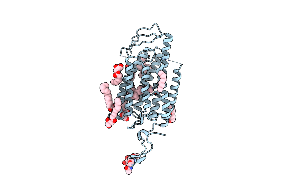
Time-Resolved Serial Femtosecond Crystallography Reveals Early Structural Changes In Channelrhodopsin: 1 Ms Structure
Organism: Chlamydomonas reinhardtii
Method: X-RAY DIFFRACTION Resolution:2.50 Å Release Date: 2021-04-07 Classification: MEMBRANE PROTEIN Ligands: RET, OLC |
Organism: Chlamydomonas reinhardtii
Method: X-RAY DIFFRACTION
Release Date: 2021-04-07
Ligands: RET, OLC
 |
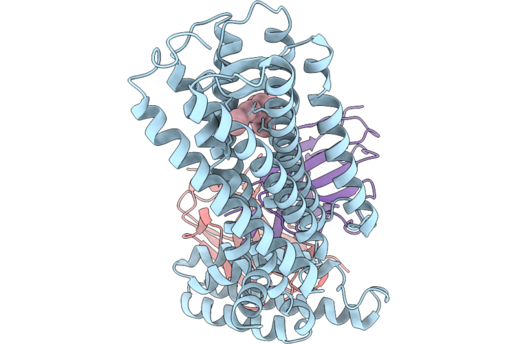
Time-Resolved Serial Femtosecond Crystallography Reveals Early Structural Changes In Channelrhodopsin: 50 Microsecond Structure
Organism: Chlamydomonas reinhardtii
Method: X-RAY DIFFRACTION Resolution:2.50 Å Release Date: 2021-04-07 Classification: MEMBRANE PROTEIN Ligands: RET, OLC |
Organism: Chlamydomonas reinhardtii
Method: X-RAY DIFFRACTION
Release Date: 2021-04-07
Ligands: RET, OLC
 |
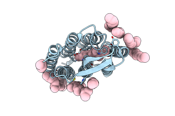
Dark-State Structure Of Human Medium-Wave-Sensitive Cone Opsin (Opn1Mw)
Organism: Homo sapiens, Escherichia coli, Synthetic construct
Method: ELECTRON MICROSCOPY Resolution:3.70 Å Release Date: 2026-06-24 Classification: MEMBRANE PROTEIN Ligands: RET |
Organism: Homo sapiens, Escherichia coli, Synthetic construct
Method: ELECTRON MICROSCOPY
Release Date: 2026-06-24
Ligands: RET
 |
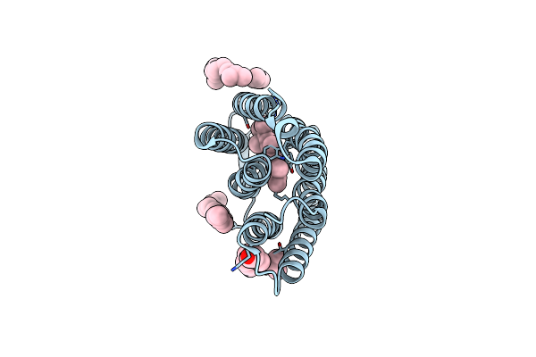
Bacteriorhodopsin Ground State Structure Collected In Cryo Conditions From Crystals Obtained In Lcp With Peg As A Precipitant.
Organism: Halobacterium salinarum
Method: X-RAY DIFFRACTION Resolution:1.90 Å Release Date: 2015-02-18 Classification: PROTON TRANSPORT Ligands: RET, LI1 |
Organism: Halobacterium salinarum
Method: X-RAY DIFFRACTION
Release Date: 2015-02-18
Ligands: RET, LI1
 |
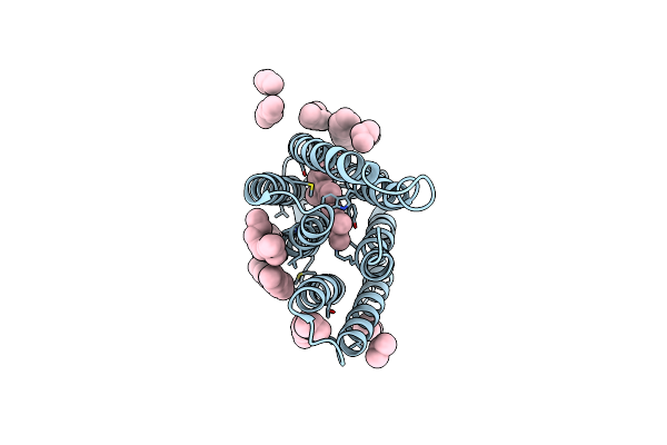
Bacteriorhodopsin Ground State Structure Obtained With Serial Femtosecond Crystallography
Organism: Halobacterium salinarum (strain atcc 700922 / jcm 11081 / nrc-1)
Method: X-RAY DIFFRACTION Resolution:2.30 Å Release Date: 2016-08-31 Classification: PROTON TRANSPORT Ligands: RET, LI1 |
Organism: Halobacterium salinarum (strain atcc 700922 / jcm 11081 / nrc-1)
Method: X-RAY DIFFRACTION
Release Date: 2016-08-31
Ligands: RET, LI1
 |
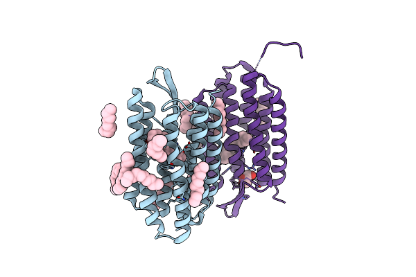
Deprotonation Of D96 In Bacteriorhodopsin Opens The Proton Uptake Pathway
Organism: Halobacterium sp. nrc-1
Method: X-RAY DIFFRACTION Resolution:2.65 Å Release Date: 2013-02-27 Classification: TRANSPORT PROTEIN Ligands: RET, CL, LI1 |
Organism: Halobacterium sp. nrc-1
Method: X-RAY DIFFRACTION
Release Date: 2013-02-27
Ligands: RET, CL, LI1
 |
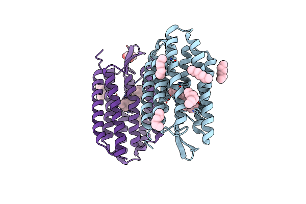
Crystal Structure Analysis Of The Bacteriorhodopsin In Facial Amphiphile-4 Dmpc Bicelle
Organism: Halobacterium salinarum
Method: X-RAY DIFFRACTION Resolution:1.99 Å Release Date: 2013-03-20 Classification: PROTON TRANSPORT Ligands: RET, OCT, D10, HEX, C14, GOL |
Organism: Halobacterium salinarum
Method: X-RAY DIFFRACTION
Release Date: 2013-03-20
Ligands: RET, OCT, D10, HEX, C14, GOL
 |
Crystal Structure Analysis Of The Bacteriorhodopsin In Facial Amphiphile-7 Dmpc Bicelle
Organism: Halobacterium salinarum
Method: X-RAY DIFFRACTION Resolution:2.00 Å Release Date: 2013-03-20 Classification: PROTON TRANSPORT Ligands: RET, HEX, HP6, GOL |
Organism: Halobacterium salinarum
Method: X-RAY DIFFRACTION
Release Date: 2013-03-20
Ligands: RET, HEX, HP6, GOL