

Search Count: 6,259
 |
Grimophan Synthase From Variovorax Boronicumulans (Gris): Open Conformation
Organism: Variovorax boronicumulans
Method: X-RAY DIFFRACTION Resolution:2.40 Å Release Date: 2026-09-30 Classification: LYASE Ligands: PO4, PEG, EDO |
Organism: Variovorax boronicumulans
Method: X-RAY DIFFRACTION
Release Date: 2026-09-30
Ligands: PO4, PEG, EDO
 |
Chlororaphen Synthase From Pseudomonas Chlororaphis (Pc-Chlos) In Complex With Pyrophosphate
Organism: Pseudomonas chlororaphis
Method: X-RAY DIFFRACTION Resolution:2.00 Å Release Date: 2026-09-30 Classification: LYASE Ligands: MG, POP, GOL, BU3, EDO, ACT, NA |
Organism: Pseudomonas chlororaphis
Method: X-RAY DIFFRACTION
Release Date: 2026-09-30
Ligands: MG, POP, GOL, BU3, EDO, ACT, NA
 |
Chlororaphen Synthase From Pseudomonas Chlororaphis (Pc-Chlos) In Complex With Thiopyrophosphate And Bistris-Buffer
Organism: Pseudomonas chlororaphis
Method: X-RAY DIFFRACTION Resolution:1.25 Å Release Date: 2026-09-30 Classification: LYASE Ligands: MG, PIS, BTB, BU3, PEG, NA, EDO |
Organism: Pseudomonas chlororaphis
Method: X-RAY DIFFRACTION
Release Date: 2026-09-30
Ligands: MG, PIS, BTB, BU3, PEG, NA, EDO
 |
Chlororaphen Synthase From Pseudomonas Chlororaphis (Pc-Chlos) In Complex With Prechlororaphen Thiolopyrophosphate (A-Pcspp)
Organism: Pseudomonas chlororaphis
Method: X-RAY DIFFRACTION Resolution:1.90 Å Release Date: 2026-09-30 Classification: LYASE Ligands: MG, A1KN5, PEG, BU3, EDO, ACT, TRS, NA |
Organism: Pseudomonas chlororaphis
Method: X-RAY DIFFRACTION
Release Date: 2026-09-30
Ligands: MG, A1KN5, PEG, BU3, EDO, ACT, TRS, NA
 |
Apo Pdz Domain From Mouse Shank3 (570-664)
Organism: Mus musculus
Method: X-RAY DIFFRACTION Resolution:1.26 Å Release Date: 2026-09-30 Classification: PROTEIN BINDING Ligands: GOL |
Organism: Mus musculus
Method: X-RAY DIFFRACTION
Release Date: 2026-09-30
Ligands: GOL
 |
Bacteroides Fragilis Carboxyaminopropylagmatine Dehydrogenase - Apo
Organism: Bacteroides fragilis cl05t12c13
Method: X-RAY DIFFRACTION Resolution:2.91 Å Release Date: 2026-09-30 Classification: BIOSYNTHETIC PROTEIN Ligands: K, GOL |
Organism: Bacteroides fragilis cl05t12c13
Method: X-RAY DIFFRACTION
Release Date: 2026-09-30
Ligands: K, GOL
 |
Cyp3A4 Cryoem Structure
Organism: Homo sapiens
Method: ELECTRON MICROSCOPY Release Date: 2026-09-30 Classification: OXIDOREDUCTASE Ligands: HEM |
Organism: Homo sapiens
Method: ELECTRON MICROSCOPY
Release Date: 2026-09-30
Ligands: HEM
 |
Cyp3A4 Bound To Ritonavir
Organism: Homo sapiens
Method: ELECTRON MICROSCOPY Release Date: 2026-09-30 Classification: OXIDOREDUCTASE Ligands: HEM, RIT |
Organism: Homo sapiens
Method: ELECTRON MICROSCOPY
Release Date: 2026-09-30
Ligands: HEM, RIT
 |
Cyp3A4 Bound To Ketoconazole
Organism: Homo sapiens
Method: ELECTRON MICROSCOPY Release Date: 2026-09-30 Classification: OXIDOREDUCTASE Ligands: HEM, KLN, KKK |
Organism: Homo sapiens
Method: ELECTRON MICROSCOPY
Release Date: 2026-09-30
Ligands: HEM, KLN, KKK
 |
Cyp3A4 Bound To Azamulin
Organism: Homo sapiens
Method: ELECTRON MICROSCOPY Release Date: 2026-09-30 Classification: OXIDOREDUCTASE Ligands: HEM, MWY |
Organism: Homo sapiens
Method: ELECTRON MICROSCOPY
Release Date: 2026-09-30
Ligands: HEM, MWY
 |
Cyp3A4 Bound To Vardenafil
Organism: Homo sapiens
Method: ELECTRON MICROSCOPY Resolution:3.50 Å Release Date: 2026-09-30 Classification: OXIDOREDUCTASE Ligands: VDN, HEM |
Organism: Homo sapiens
Method: ELECTRON MICROSCOPY
Release Date: 2026-09-30
Ligands: VDN, HEM
 |
Clostridium Leptum Carboxyaminopropylagmatine Dehydrogenase Apo
Organism: [clostridium] leptum
Method: X-RAY DIFFRACTION Resolution:2.50 Å Release Date: 2026-09-30 Classification: OXIDOREDUCTASE Ligands: GOL |
Organism: [clostridium] leptum
Method: X-RAY DIFFRACTION
Release Date: 2026-09-30
Ligands: GOL
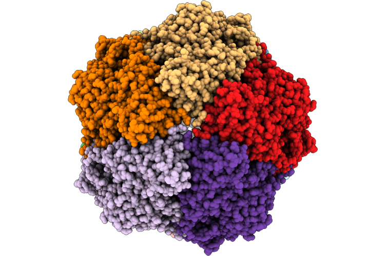 |
Zuzalysin Bi-Pentamer Cryo-Em
Organism: Porphyromonas gingivalis
Method: ELECTRON MICROSCOPY Release Date: 2026-09-23 Classification: HYDROLASE Ligands: CA, ZN |
Organism: Porphyromonas gingivalis
Method: ELECTRON MICROSCOPY
Release Date: 2026-09-23
Ligands: CA, ZN
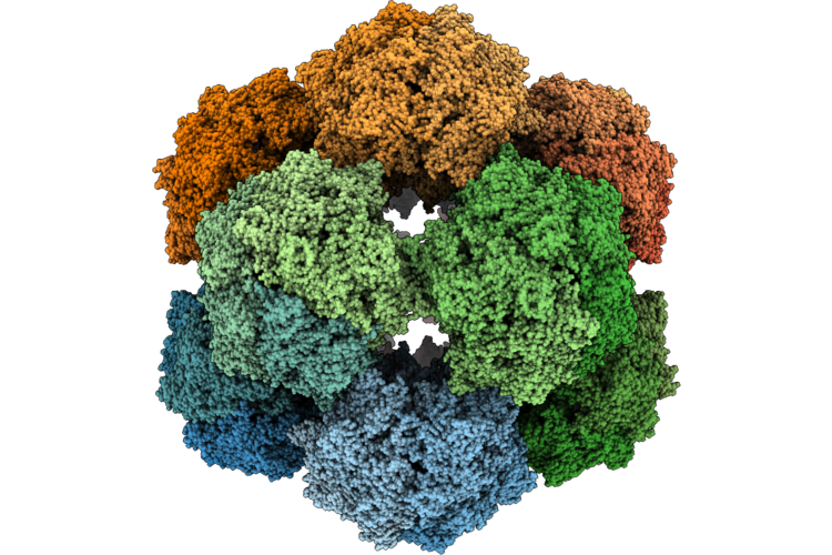 |
Zuzalysin Zymogen Dodecahedral Complex E439A
Organism: Porphyromonas gingivalis
Method: ELECTRON MICROSCOPY Release Date: 2026-09-23 Classification: HYDROLASE Ligands: CA, ZN |
Organism: Porphyromonas gingivalis
Method: ELECTRON MICROSCOPY
Release Date: 2026-09-23
Ligands: CA, ZN
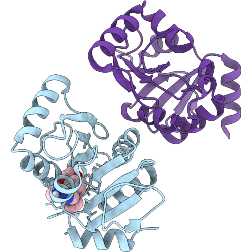 |
Pandda Analysis Group Deposition -- Crystal Structure Of Sars-Cov-2 Nsp3 Macrodomain In Complex With Z1039063794
Organism: Severe acute respiratory syndrome coronavirus 2
Method: X-RAY DIFFRACTION Resolution:0.98 Å Release Date: 2026-09-16 Classification: VIRAL PROTEIN Ligands: A1DBS |
Organism: Severe acute respiratory syndrome coronavirus 2
Method: X-RAY DIFFRACTION
Release Date: 2026-09-16
Ligands: A1DBS
 |
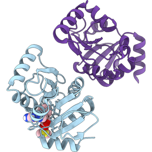
Pandda Analysis Group Deposition -- Crystal Structure Of Sars-Cov-2 Nsp3 Macrodomain In Complex With Z7237399929
Organism: Severe acute respiratory syndrome coronavirus 2
Method: X-RAY DIFFRACTION Resolution:1.01 Å Release Date: 2026-09-16 Classification: VIRAL PROTEIN Ligands: A1DBO, A1CJ0 |
Organism: Severe acute respiratory syndrome coronavirus 2
Method: X-RAY DIFFRACTION
Release Date: 2026-09-16
Ligands: A1DBO, A1CJ0
 |
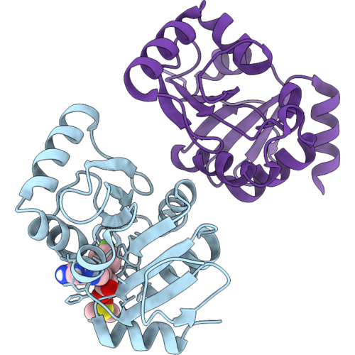
Pandda Analysis Group Deposition -- Crystal Structure Of Sars-Cov-2 Nsp3 Macrodomain In Complex With Z7534232254
Organism: Severe acute respiratory syndrome coronavirus 2
Method: X-RAY DIFFRACTION Resolution:1.01 Å Release Date: 2026-09-16 Classification: VIRAL PROTEIN Ligands: DMS, A1DBT, A1CKL |
Organism: Severe acute respiratory syndrome coronavirus 2
Method: X-RAY DIFFRACTION
Release Date: 2026-09-16
Ligands: DMS, A1DBT, A1CKL
 |
Pandda Analysis Group Deposition -- Crystal Structure Of Sars-Cov-2 Nsp3 Macrodomain In Complex With Z7691912478
Organism: Severe acute respiratory syndrome coronavirus 2
Method: X-RAY DIFFRACTION Resolution:0.97 Å Release Date: 2026-09-16 Classification: VIRAL PROTEIN Ligands: DMS, A1AKF, A1CKA |
Organism: Severe acute respiratory syndrome coronavirus 2
Method: X-RAY DIFFRACTION
Release Date: 2026-09-16
Ligands: DMS, A1AKF, A1CKA
 |
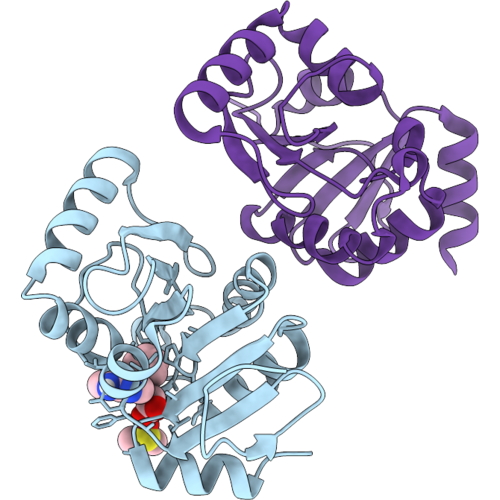
Pandda Analysis Group Deposition -- Crystal Structure Of Sars-Cov-2 Nsp3 Macrodomain In Complex With Z7983841111
Organism: Severe acute respiratory syndrome coronavirus 2
Method: X-RAY DIFFRACTION Resolution:1.00 Å Release Date: 2026-09-16 Classification: VIRAL PROTEIN Ligands: A1DBU, A1CKC |
Organism: Severe acute respiratory syndrome coronavirus 2
Method: X-RAY DIFFRACTION
Release Date: 2026-09-16
Ligands: A1DBU, A1CKC
 |
Pandda Analysis Group Deposition -- Crystal Structure Of Sars-Cov-2 Nsp3 Macrodomain In Complex With Z8727396600
Organism: Severe acute respiratory syndrome coronavirus 2
Method: X-RAY DIFFRACTION Resolution:0.97 Å Release Date: 2026-09-16 Classification: VIRAL PROTEIN Ligands: DMS, A1DBV |
Organism: Severe acute respiratory syndrome coronavirus 2
Method: X-RAY DIFFRACTION
Release Date: 2026-09-16
Ligands: DMS, A1DBV