9YC5 image
Deposition Date 2025-09-18
Release Date 2026-02-04
Last Version Date 2026-02-04
Entry Detail
PDB ID:
9YC5
Keywords:
Title:
Human uPAR bound to the Fab fragment of targeted cancer therapeutic antibody FL1
Biological Source:
Source Organism(s):
Homo sapiens (Taxon ID: 9606)
Mus musculus (Taxon ID: 10090)
Expression System(s):
Method Details:
Experimental Method:
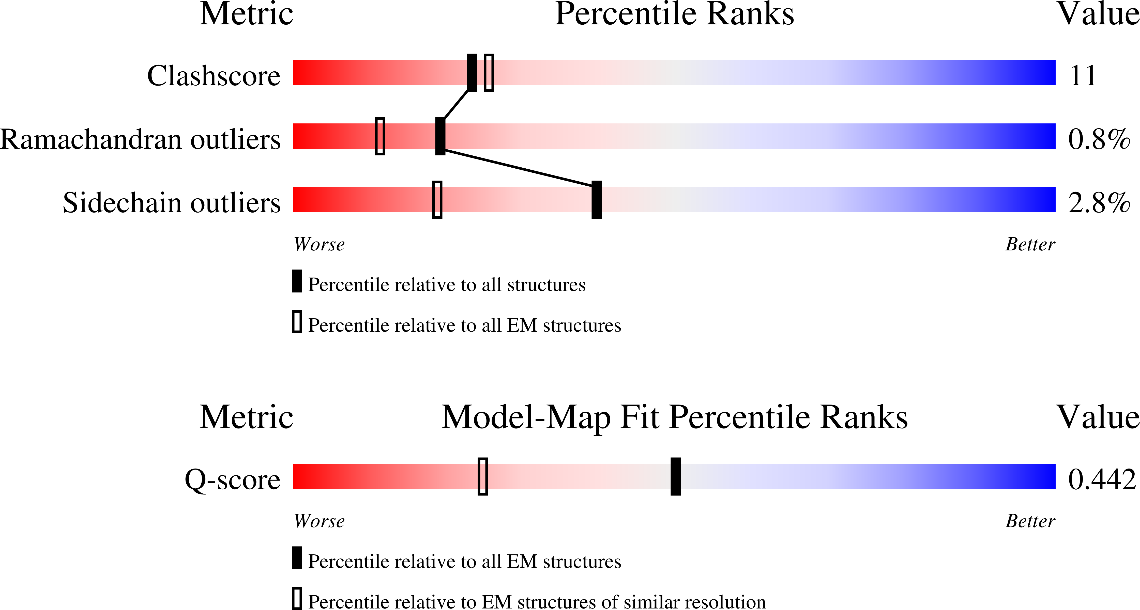
Resolution:
2.94 Å
Aggregation State:
PARTICLE
Reconstruction Method:
SINGLE PARTICLE
Macromolecular Entities
Structures with similar UniProt ID
Protein Blast
Polymer Type:polypeptide(L)
Molecule:Urokinase plasminogen activat
Gene (Uniprot):PLAUR
Chain IDs:A
Chain Length:283
Number of Molecules:1
Biological Source:Homo sapiens
Polymer Type:polypeptide(L)
Molecule:Anti-uPAR antibody FL1 Fab he
Chain IDs:B (auth: H)
Chain Length:221
Number of Molecules:1
Biological Source:Mus musculus
Polymer Type:polypeptide(L)
Molecule:Anti-uPAR antibody FL1 Fab li
Chain IDs:C (auth: L)
Chain Length:219
Number of Molecules:1
Biological Source:Mus musculus
Ligand Molecules
Primary Citation
Structural basis for uPAR binding to an antibody developed for targeted cancer therapy. Mechanistic insights into flexibility, ligand recognition, and molecular imaging.
Protein Sci. 35 e70473 e70473 (2026)
PMID: 41575054 DOI: 10.1002/pro.70473

Abstact

The urokinase-type plasminogen activator receptor (uPAR) is currently gaining momentum as a promising molecular target for treatment of various solid cancers. For patient stratification, we developed a high-affinity uPAR-targeting peptide (AE105) detecting primary cancer lesions as well as occult metastasis by positron emission tomography (PET) imaging. uPAR-targeting by AE105 is also used for optical imaging in fluorescence-guided surgery of, for example, head-and-neck cancers. Recently, we showed that a monoclonal anti-uPAR antibody (FL1), in the form of an antibody-drug conjugate (FL1-ADC), efficiently eradicate pancreatic ductal carcinomas in surrogate mouse models leading to long-term remissions. In the current study, we solved high-resolution cryo-EM structures of FL1 in complex with two different conformational states of uPAR. Combined with comprehensive kinetic data from surface plasmon resonance studies, our cryo-EM structures provide essential insights into how FL1 binding impacts the interdomain flexibility of uPAR by restricting the movement of its N-terminal LU domain. This constraint from the bound FL1 drives uPAR into its open conformation, which leads to a pronounced reduction in the binding affinity for both its natural protease ligand (300-fold) and the PET imaging probe AE105 (25-fold). Collectively, these consequences of FL1-binding on uPAR conformation are considered beneficial for both targeted cancer treatment with FL1-ADCs and for the accompanying evaluation of treatment efficacy by longitudinal AE105-based PET imaging.

Legend

Protein

Chemical

Disease

Primary Citation of related structures
Feedback Form
Name
Email
Institute
Feedback