9P0V image
Deposition Date 2025-06-07
Release Date 2026-02-04
Last Version Date 2026-03-25
Entry Detail
PDB ID:
9P0V
Title:
Structure of PYCR1 complexed with the allosteric inhibitor 7-benzamido-4-hydroxynaphthalene-2-sulfonic acid in a remote site and NADH and (S)-(-)-2-Hydroxy-3,3-dimethylbutyric acid in the active site
Biological Source:
Source Organism(s):
Homo sapiens (Taxon ID: 9606)
Expression System(s):
Method Details:
Experimental Method:
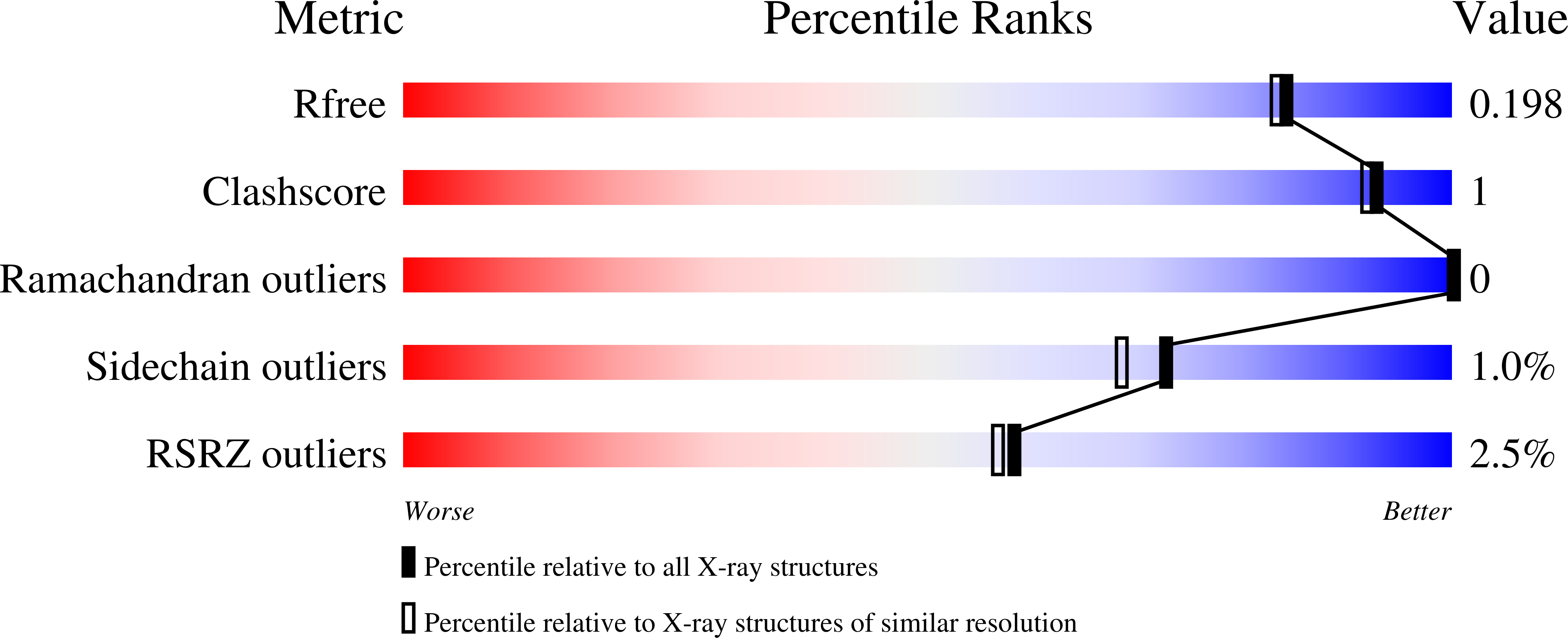
Resolution:
1.80 Å
R-Value Free:
0.20
R-Value Work:
0.17
R-Value Observed:
0.17
Space Group:
C 1 2 1
Macromolecular Entities
Structures with similar UniProt ID
Protein Blast
Polymer Type:polypeptide(L)
Molecule:Pyrroline-5-carboxylate reduc
Gene (Uniprot):PYCR1
Chain IDs:A, B, C, D, E
Chain Length:316
Number of Molecules:5
Biological Source:Homo sapiens
Primary Citation
Serendipitous discovery of an allosteric inhibitor binding groove in the proline biosynthetic enzyme pyrroline-5-carboxylate reductase 1.
Biochem.J. 483 409 427 (2026)
PMID: 41569420 DOI: 10.1042/BCJ20250278

Abstact

Δ1-pyrroline-5-carboxylate (P5C) reductase 1 (PYCR1) catalyzes the NAD(P)H-dependent conversion of L-P5C to L-proline and is one of the most consistently upregulated metabolic enzymes in cancer cells. High PYCR1 expression is associated with adverse clinical outcomes, and its knockdown inhibits tumor proliferation and metastasis, motivating inhibitor discovery. All structurally validated PYCR1 inhibitors to date bind in the active site and are anchored in the L-P5C binding pocket by an anionic functional group, typically carboxylate. Seeking inhibitors with alternative anchors, we used X-ray crystallography to screen 22 fragment-like compounds (MW = 189-343 Da) from docking that represent six different carboxylic acid isosteres. Surprisingly, only one compound bound in the active site. Four other compounds were found in three adjacent remote sites located in oligomer interfaces. The compounds bind 7 Å from NADH and 10-14 Å from L-P5C, and the intervening space is blocked by protein for inhibitors in Sites 1A/1B and open for inhibitors in Site 2. Together, the three binding sites define a ligand binding hot spot groove that spans 33 Å. The remote binders inhibit PYCR1 activity with K values from the mixed model of inhibition of 32 μM to 2 mM. Co-crystal structures of PYCR1 with combinations of allosteric inhibitors, NADH, and L-P5C/proline analogs suggest the inhibitors can bind to the ternary PYCR1-L-P5C-NAD(P)H complex in addition to the free enzyme, consistent with a mixed mechanism of inhibition. The discovery of an allosteric inhibitor binding groove that accommodates multiple fragments heralds a new era of PYCR1 inhibitor design.

Legend

Protein

Chemical

Disease

Primary Citation of related structures
Feedback Form
Name
Email
Institute
Feedback