9O5G image
Deposition Date 2025-04-10
Release Date 2026-02-04
Last Version Date 2026-02-04
Entry Detail
PDB ID:
9O5G
Keywords:
Title:
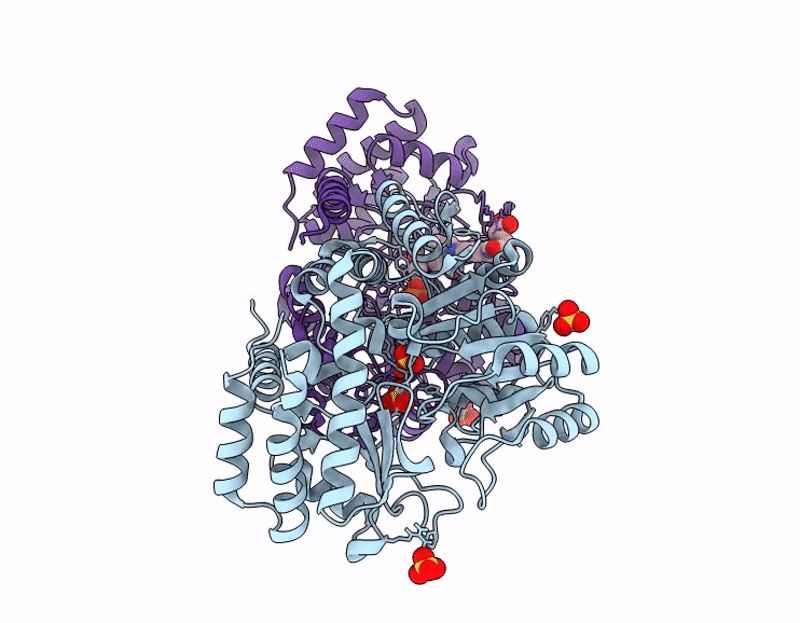
Room-temperature joint X-ray/Neutron structure of Thermus thermophilus SHMT in complex with PLP-Gly external aldimine and 5-methyl-tetrahydrofolate (5MTHF)
Biological Source:
Source Organism(s):
Expression System(s):
Method Details:
R-Value Free:
['0.32
R-Value Work:
['0.29
R-Value Observed:
['?', '?'].00
Space Group:
P 1 21 1
Macromolecular Entities
Structures with similar UniProt ID
Protein Blast
Polymer Type:polypeptide(L)
Molecule:Serine hydroxymethyltransfera
Gene (Uniprot):glyA
Chain IDs:A, B
Chain Length:405
Number of Molecules:2
Biological Source:Thermus thermophilus
Primary Citation
Neutron diffraction reveals protonation states in pyridoxal-5'-phosphate-free and glycine external aldimine-bound serine hydroxymethyltransferase.
Febs J. 293 582 597 (2026)
PMID: 40915980 DOI: 10.1111/febs.70260

Abstact

Serine hydroxymethyltransferase (SHMT) is a critical enzyme in the one-carbon (1C) metabolism pathway catalyzing the reversible conversion of L-Ser into Gly and concurrent transfer of 1C unit to tetrahydrofolate (THF) to give 5,10-methylene-THF (5,10-MTHF), which is used in the downstream syntheses of biomolecules critical for cell proliferation. The cellular 1C metabolism is hijacked by many cancer types to support cancer cell proliferation, making SHMT a promising target for the design and development of novel small-molecule antimetabolite chemotherapies. To advance structure-assisted drug design, knowledge of SHMT catalysis is crucial, but can only be fully realized when the atomic details of each reaction step governed by the acid-base catalysis are elucidated by visualizing active site hydrogen atoms. Here, we used room-temperature neutron crystallography to directly determine protonation states in Thermus thermophilus SHMT (TthSHMT), capturing protomer A in the apo form lacking the coenzyme pyridoxal 5'-phosphate (PLP), and protomer B as a ternary complex with PLP-Gly-external aldimine and (6S)-5-methyltetrahydrofolate (5MTHF). We observed protonation of the Schiff base nitrogen in PLP-Gly and neutrality of the catalytic Lys226 side chain in the ternary complex, whereas Lys226 is protonated and positively charged in the apo-active site. Furthermore, we obtained an X-ray structure of TthSHMT in complex with the substrate THF, which binds identically as 5MTHF at the peripheral binding site. The unique structural and functional information provided by neutron crystallography, in combination with X-ray structures, can be employed in the rational design of SHMT inhibitors.

Legend

Protein

Chemical

Disease

Primary Citation of related structures
Feedback Form
Name
Email
Institute
Feedback