9NX2 image
Deposition Date 2025-03-25
Release Date 2026-02-04
Last Version Date 2026-03-25
Entry Detail
PDB ID:
9NX2
Title:
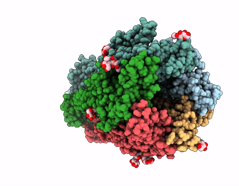
Muscle-type nicotinic acetylcholine receptor bound to conotoxin ImII
Biological Source:
Source Organism(s):
Method Details:
Experimental Method:
Resolution:
2.96 Å
Aggregation State:
PARTICLE
Reconstruction Method:
SINGLE PARTICLE
Macromolecular Entities
Structures with similar UniProt ID
Protein Blast
Polymer Type:polypeptide(L)
Molecule:Acetylcholine receptor subuni
Gene (Uniprot):CHRNA1
Chain IDs:A, D
Chain Length:434
Number of Molecules:2
Biological Source:Tetronarce californica
Structures with similar UniProt ID
Protein Blast
Polymer Type:polypeptide(L)
Molecule:Acetylcholine receptor subuni
Gene (Uniprot):chrnd
Chain IDs:B
Chain Length:499
Number of Molecules:1
Biological Source:Tetronarce californica
Structures with similar UniProt ID
Protein Blast
Polymer Type:polypeptide(L)
Molecule:Acetylcholine receptor subuni
Gene (Uniprot):CHRNB1
Chain IDs:C
Chain Length:469
Number of Molecules:1
Biological Source:Tetronarce californica
Structures with similar UniProt ID
Protein Blast
Polymer Type:polypeptide(L)
Molecule:Acetylcholine receptor subuni
Gene (Uniprot):CHRNG
Chain IDs:E
Chain Length:489
Number of Molecules:1
Biological Source:Tetronarce californica
Structures with similar UniProt ID
Protein Blast
Polymer Type:polypeptide(L)
Molecule:Alpha-conotoxin ImII
Chain IDs:F, G
Chain Length:13
Number of Molecules:2
Biological Source:Conus imperialis
Primary Citation
Shape-shifting conotoxins reveal divergent pore-targeting mechanisms in nicotinic receptors.
Structure 34 463 ? (2026)
PMID: 41468893 DOI: 10.1016/j.str.2025.12.003

Abstact

The neuronal α7 nicotinic acetylcholine receptor (α7-nAChR) and muscle-type nicotinic acetylcholine receptor (mt-nAChR) are pivotal in synaptic signaling within the brain and the neuromuscular junction respectively. Additionally, they are both targets of a wide range of drugs and toxins. Here, we utilize cryo-EM to delineate structures of these nAChRs in complex with the conotoxins ImI and ImII from Conus imperialis. Despite nominal sequence differences, ImI and ImII exhibit discrete binding preferences and adopt drastically different conformational states upon binding. ImI engages the orthosteric sites of α7-nAChR, while ImII forms distinct pore-bound complexes with both α7-nAChR and mt-nAChR. Strikingly, ImII adopts a compact globular conformation that binds as a monomer to the α7-nAChR pore and as an oblate dimer to the mt-nAChR pore. These structures advance our understanding of nAChR-ligand interactions and the subtle sequence variations that result in dramatically altered functional outcomes in small peptide toxins.

Legend

Protein

Chemical

Disease

Primary Citation of related structures
Feedback Form
Name
Email
Institute
Feedback