9MVG image
Deposition Date 2025-01-15
Release Date 2026-01-28
Last Version Date 2026-02-18
Entry Detail
PDB ID:
9MVG
Keywords:
Title:
Structure of SciW variant L66A bound to the Rhs1 transmembrane domain
Biological Source:
Method Details:
Experimental Method:
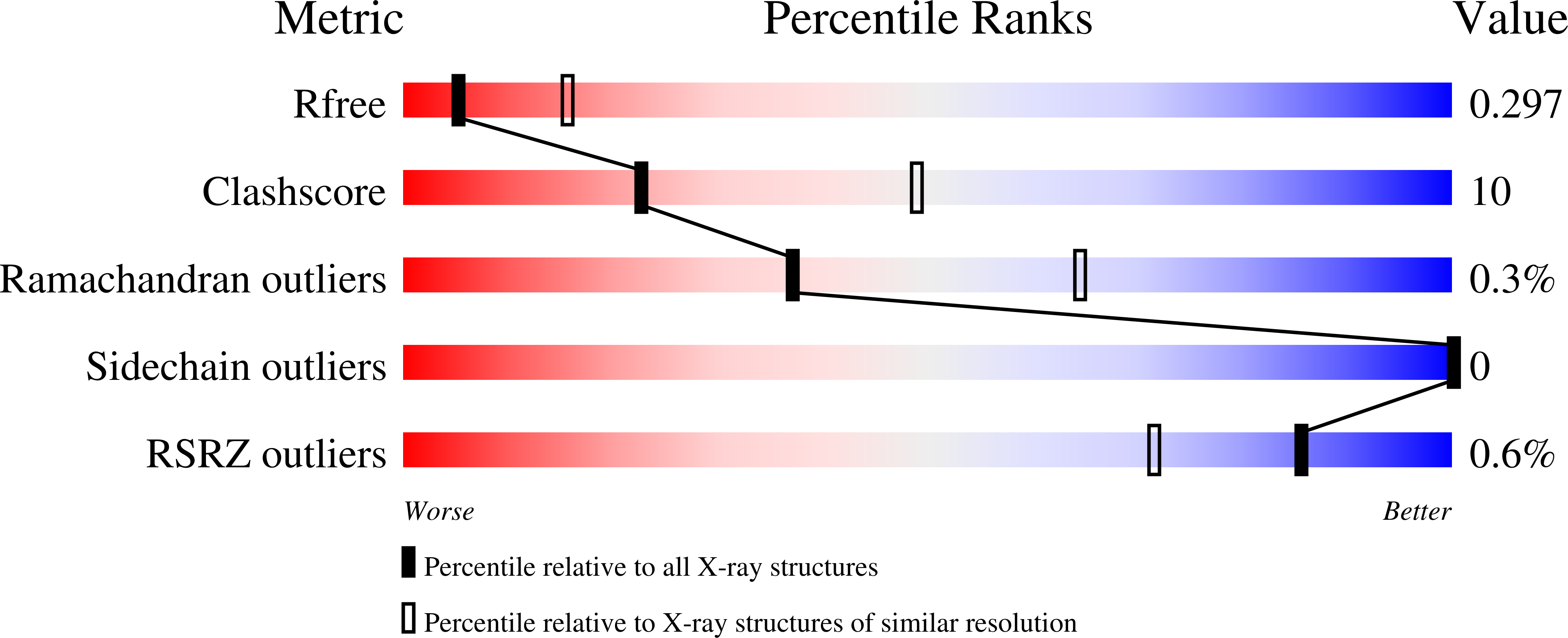
Resolution:
3.05 Å
R-Value Free:
0.29
R-Value Work:
0.26
R-Value Observed:
0.26
Space Group:
C 1 2 1
Macromolecular Entities
Structures with similar UniProt ID
Protein Blast
Polymer Type:polypeptide(L)
Molecule:SciW protein
Gene (Uniprot):sciw
Mutagens:L66A
Chain IDs:A, B, D, E
Chain Length:148
Number of Molecules:4
Biological Source:Salmonella enterica subsp. enterica serovar Typhimurium
Structures with similar UniProt ID
Protein Blast
Polymer Type:polypeptide(L)
Molecule:Rhs1 protein
Gene (Uniprot):G0K95_003520
Chain IDs:C, F
Chain Length:66
Number of Molecules:2
Biological Source:Salmonella enterica subsp. enterica serovar Typhimurium
Primary Citation
Biophysical characterization of Eag chaperones suggests the mechanism of effector transmembrane domain release.
Nat Commun 17 1401 1401 (2026)
PMID: 41571669 DOI: 10.1038/s41467-025-68138-w

Abstact

The type VI secretion system (T6SS) is a dynamic protein nanomachine found in Gram-negative bacteria that secretes toxic effectors into prey-cells. For secretion, effectors require chaperones or adaptors for proper loading onto the T6SS. Effector associated genes (Eags) are a family of T6SS chaperones that stabilize N-terminal transmembrane domains (TMDs) found in thousands of effectors. Eags are essential for secretion and inhibit effector TMDs from prematurely adopting a membrane-penetrative conformation. However, the mechanism of TMD release from its cognate Eag chaperone is unknown. Here, we take a biochemical and biophysical approach to probe the mechanism of TMD binding and dissociation from Eag chaperones. Using steady-state fluorescence, stopped-flow measurements, and bacterial competition assays, we compare the thermodynamics, kinetics, and in vivo chaperone function of wild-type and point variant Eag-TMD complexes. Additionally, we solve an X-ray crystal structure of an Eag-TMD point variant complex that captures an intermediate state of TMD release. Our data reveals the molecular features and specific residue contacts necessary for TMD binding and demonstrates the Eag conformational change required to initiate rapid release of the TMD. Overall, our work details the stability of Eag-TMD complexes and the energetic pathway for the dissociation of effector TMDs from their Eag chaperones.

Legend

Protein

Chemical

Disease

Primary Citation of related structures
Feedback Form
Name
Email
Institute
Feedback