9EDT image
Deposition Date 2024-11-18
Release Date 2026-02-04
Last Version Date 2026-02-18
Entry Detail
PDB ID:
9EDT
Title:
Tubulin cofactors D,E,G bound to tubulin dimer
Biological Source:
Source Organism(s):
Expression System(s):
Method Details:
Experimental Method:
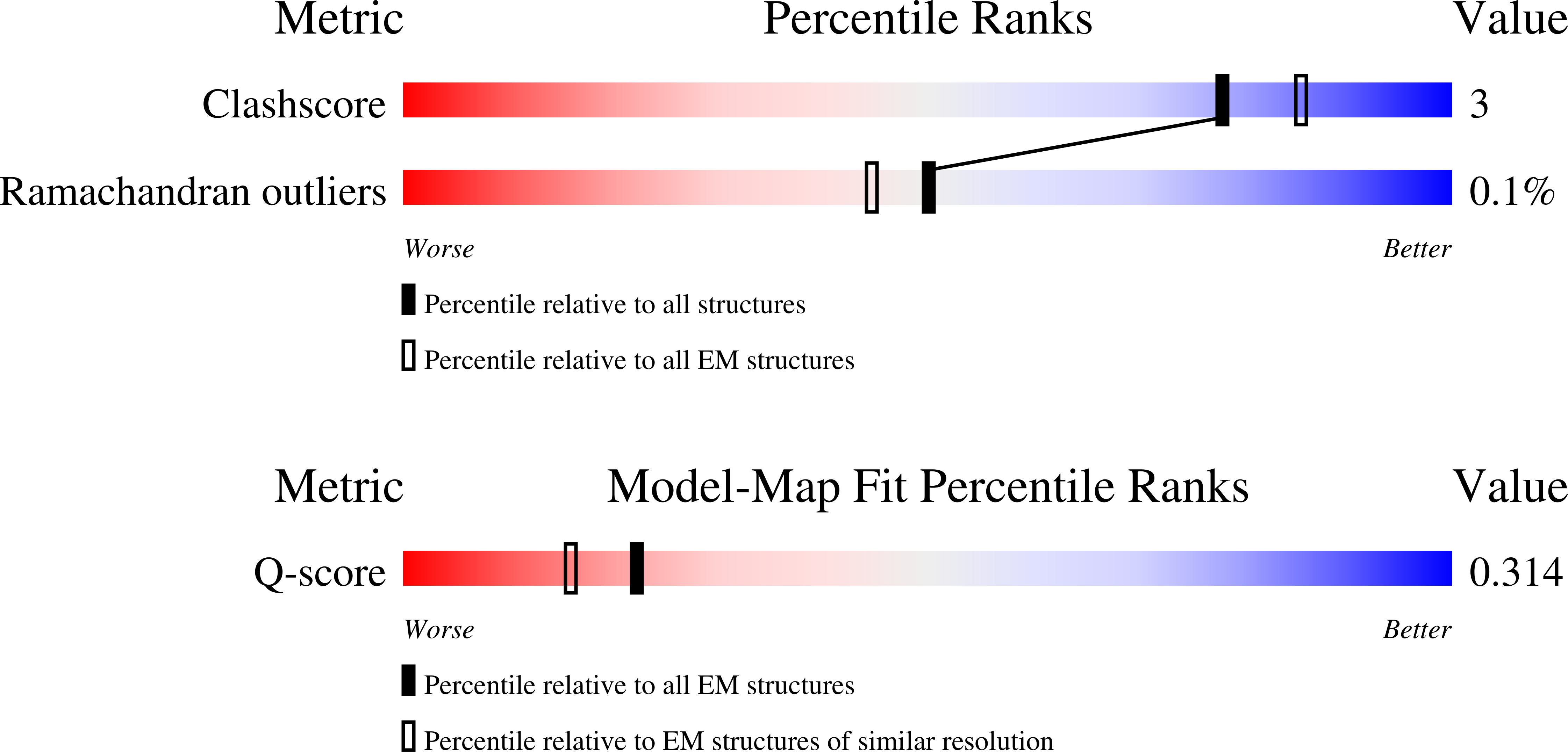
Resolution:
3.70 Å
Aggregation State:
PARTICLE
Reconstruction Method:
SINGLE PARTICLE
Macromolecular Entities
Structures with similar UniProt ID
Protein Blast
Polymer Type:polypeptide(L)
Molecule:Tubulin alpha-1A chain
Gene (Uniprot):TUBA1A
Chain IDs:C (auth: A)
Chain Length:451
Number of Molecules:1
Biological Source:Sus scrofa
Structures with similar UniProt ID
Protein Blast
Polymer Type:polypeptide(L)
Molecule:Tubulin beta chain
Chain IDs:D (auth: B)
Chain Length:445
Number of Molecules:1
Biological Source:Sus scrofa
Structures with similar UniProt ID
Protein Blast
Polymer Type:polypeptide(L)
Molecule:Chromosome instability protei
Gene (Uniprot):CIN1
Chain IDs:A (auth: D)
Chain Length:1020
Number of Molecules:1
Biological Source:Saccharomyces cerevisiae
Structures with similar UniProt ID
Protein Blast
Polymer Type:polypeptide(L)
Molecule:Protein PAC2
Gene (Uniprot):PAC2
Chain IDs:F (auth: E)
Chain Length:459
Number of Molecules:1
Biological Source:Saccharomyces cerevisiae
Polymer Type:polypeptide(L)
Molecule:Designed ankyrin repeat prote
Chain IDs:E (auth: F)
Chain Length:169
Number of Molecules:1
Biological Source:Escherichia coli
Structures with similar UniProt ID
Protein Blast
Polymer Type:polypeptide(L)
Molecule:GTP-binding protein CIN4
Gene (Uniprot):CIN4
Chain IDs:B (auth: G)
Chain Length:191
Number of Molecules:1
Biological Source:Saccharomyces cerevisiae
Primary Citation
Cryo-EM structures of the tubulin cofactors reveal the molecular basis of alpha/beta-tubulin biogenesis.
Nat Commun 17 1405 1405 (2025)
PMID: 41461644 DOI: 10.1038/s41467-025-68142-0

Abstact

Microtubule polarity and dynamic polymerization arise from the self-association properties of the αβ-tubulin heterodimer. For decades, it has remained unclear how the tubulin cofactors TBCD, TBCE, TBCC, and the Arl2 GTPase mediate the biogenesis of αβ-tubulin from individual α- and β-tubulins. Here, we use cryo-electron microscopy to determine structures of tubulin cofactors bound to αβ-tubulin. TBCD, TBCE, and Arl2 form a heterotrimeric cage-like assembly, we term TBC-DEG, around the αβ-tubulin heterodimer. The TBC-DEG-αβ-tubulin structures show that TBC-DEG wraps around β-tubulin while TBCE extends along α-tubulin. The TBC-DEG/TBCC-αβ-tubulin structures reveal that TBCC forms multi-domain interactions with Arl2 and TBCD to engage the αβ-tubulin intradimer-interface, promoting TBCE rotation while TBCD holds β-tubulin. TBCC engages the GTP-bound Arl2, multiple sites of TBCD, and the native αβ-tubulin intradimer interface near the α-tubulin N-site GTP. Together, these structures uncover transition states for αβ-tubulin biogenesis and degradation, suggesting a vise-like, GTP-hydrolysis-dependent mechanism in which TBCC binding to TBC-DEG modulates αβ-tubulin interfaces. Our studies provide structural evidence that tubulin cofactors act as enzymatic regulators that assemble the invariant αβ-tubulin architecture. By catalyzing α- and β-tubulin biogenesis and degradation, the TBC-DEG and TBCC assemblies regulate the polymerization competency of αβ-tubulin for microtubule formation.

Legend

Protein

Chemical

Disease

Primary Citation of related structures
Feedback Form
Name
Email
Institute
Feedback