3BUK image
Deposition Date 2008-01-02
Release Date 2008-07-15
Last Version Date 2024-10-30
Entry Detail
PDB ID:
3BUK
Title:
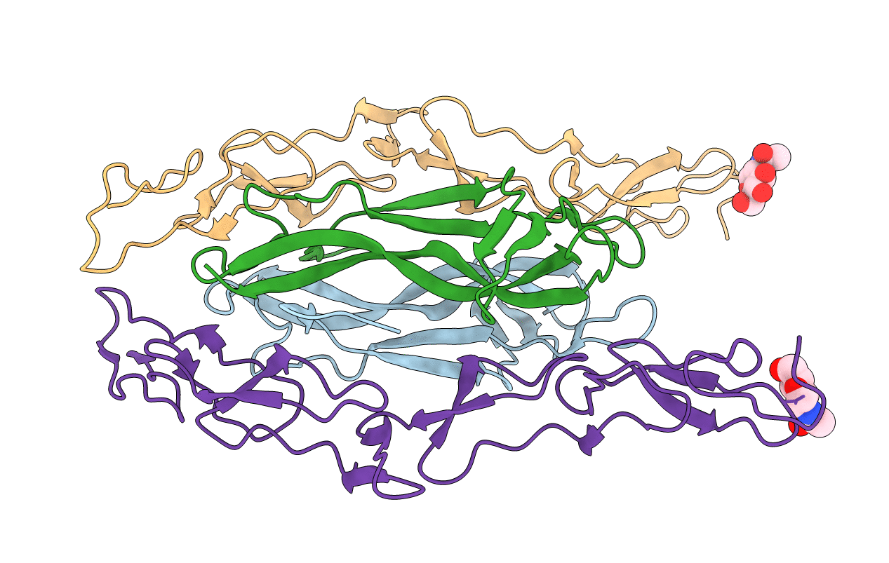
Crystal Structure of the Neurotrophin-3 and p75NTR Symmetrical Complex
Biological Source:
Source Organism(s):
Homo sapiens (Taxon ID: )
Method Details:
Experimental Method:
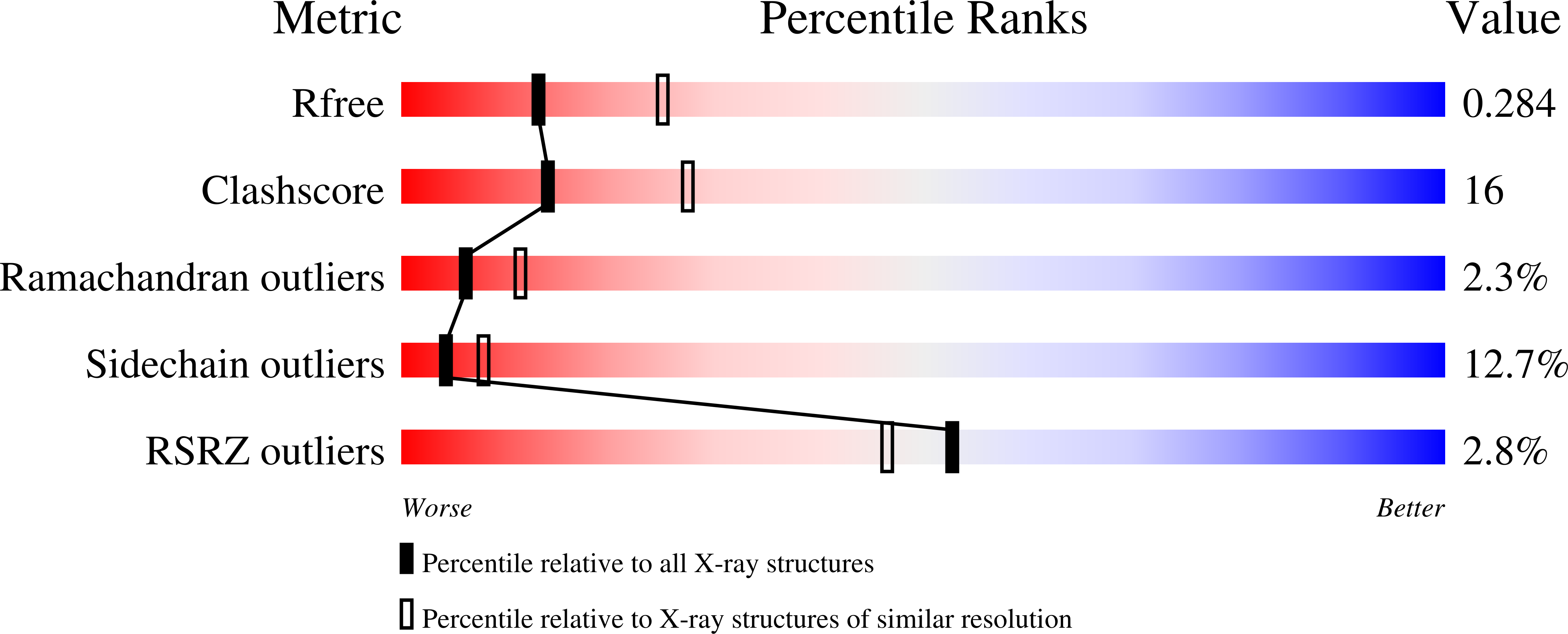
Resolution:
2.60 Å
R-Value Free:
0.28
R-Value Work:
0.22
R-Value Observed:
0.22
Space Group:
H 3
Macromolecular Entities
Structures with similar UniProt ID
Protein Blast
Polymer Type:polypeptide(L)
Molecule:Neurotrophin-3
Gene (Uniprot):NTF3
Chain IDs:A, B
Chain Length:119
Number of Molecules:2
Biological Source:Homo sapiens
Structures with similar UniProt ID
Protein Blast
Polymer Type:polypeptide(L)
Molecule:Tumor necrosis factor recepto
Gene (Uniprot):Ngfr
Chain IDs:C, D
Chain Length:167
Number of Molecules:2
Biological Source:Rattus norvegicus
Modified Residue
Compound ID Chain ID Parent Comp ID Details 2D Image
ASN C ASN GLYCOSYLATION SITE
Ligand Molecules
Primary Citation
Crystal structure of the neurotrophin-3 and p75NTR symmetrical complex.
Nature 454 789 793 (2008)
PMID: 18596692 DOI: 10.1038/nature07089

Abstact

Neurotrophins (NTs) are important regulators for the survival, differentiation and maintenance of different peripheral and central neurons. NTs bind to two distinct classes of glycosylated receptor: the p75 neurotrophin receptor (p75(NTR)) and tyrosine kinase receptors (Trks). Whereas p75(NTR) binds to all NTs, the Trk subtypes are specific for each NT. The question of whether NTs stimulate p75(NTR) by inducing receptor homodimerization is still under debate. Here we report the 2.6-A resolution crystal structure of neurotrophin-3 (NT-3) complexed to the ectodomain of glycosylated p75(NTR). In contrast to the previously reported asymmetric complex structure, which contains a dimer of nerve growth factor (NGF) bound to a single ectodomain of deglycosylated p75(NTR) (ref. 3), we show that NT-3 forms a central homodimer around which two glycosylated p75(NTR) molecules bind symmetrically. Symmetrical binding occurs along the NT-3 interfaces, resulting in a 2:2 ligand-receptor cluster. A comparison of the symmetrical and asymmetric structures reveals significant differences in ligand-receptor interactions and p75(NTR) conformations. Biochemical experiments indicate that both NT-3 and NGF bind to p75(NTR) with 2:2 stoichiometry in solution, whereas the 2:1 complexes are the result of artificial deglycosylation. We therefore propose that the symmetrical 2:2 complex reflects a native state of p75(NTR) activation at the cell surface. These results provide a model for NTs-p75(NTR) recognition and signal generation, as well as insights into coordination between p75(NTR) and Trks.

Legend

Protein

Chemical

Disease

Primary Citation of related structures
Feedback Form
Name
Email
Institute
Feedback