1OGY image
Deposition Date 2003-05-19
Release Date 2003-10-09
Last Version Date 2024-10-23
Entry Detail
PDB ID:
1OGY
Keywords:
Title:
Crystal structure of the heterodimeric nitrate reductase from Rhodobacter sphaeroides
Biological Source:
Source Organism(s):
Expression System(s):
Method Details:
Experimental Method:
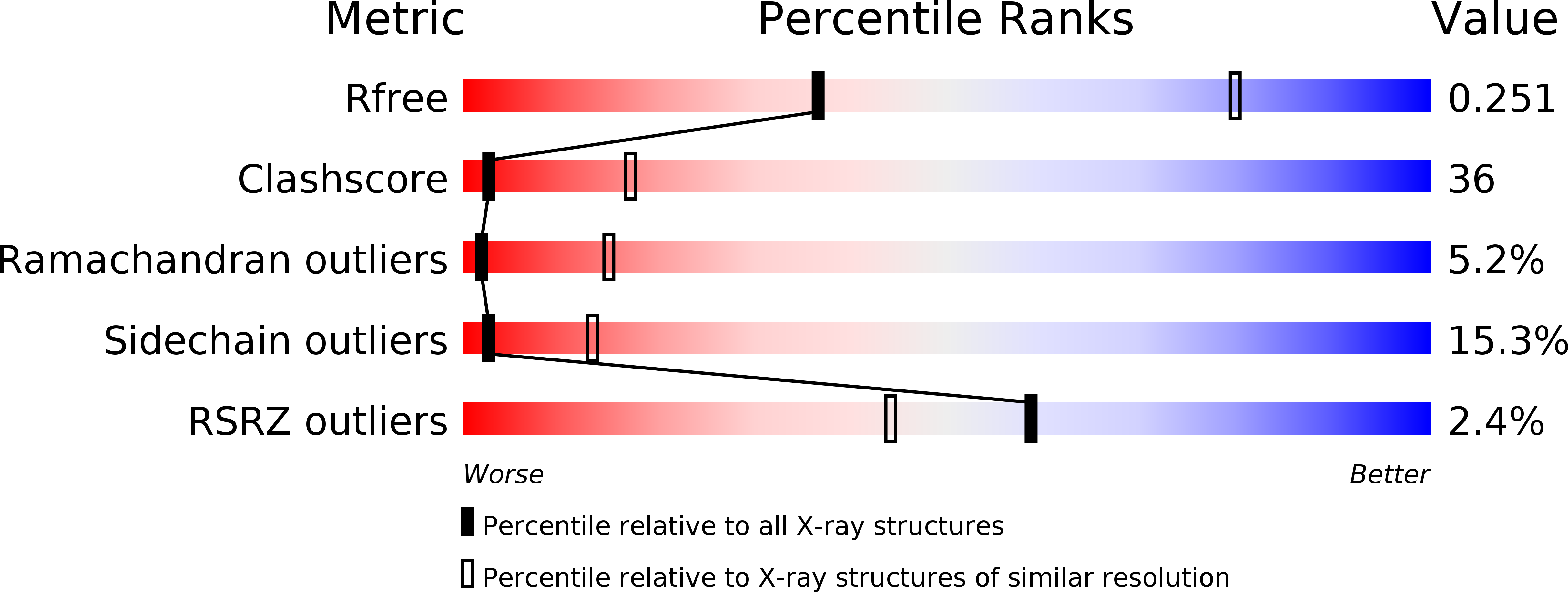
Resolution:
3.20 Å
R-Value Free:
0.26
R-Value Work:
0.25
R-Value Observed:
0.25
Space Group:
P 1 21 1
Macromolecular Entities
Structures with similar UniProt ID
Protein Blast
Polymer Type:polypeptide(L)
Molecule:PERIPLASMIC NITRATE REDUCTASE
Gene (Uniprot):napA
Chain IDs:A, C, E, G, I, K, M, O
Chain Length:802
Number of Molecules:8
Biological Source:RHODOBACTER SPHAEROIDES
Structures with similar UniProt ID
Protein Blast
Polymer Type:polypeptide(L)
Molecule:DIHEME CYTOCHROME C NAPB MOLE
Gene (Uniprot):napB
Chain IDs:B, D, F, H, J, L, N, P
Chain Length:130
Number of Molecules:8
Biological Source:RHODOBACTER SPHAEROIDES
Primary Citation

Abstact

The structure of the respiratory nitrate reductase (NapAB) from Rhodobacter sphaeroides, the periplasmic heterodimeric enzyme responsible for the first step in the denitrification process, has been determined at a resolution of 3.2 A. The di-heme electron transfer small subunit NapB binds to the large subunit with heme II in close proximity to the [4Fe-4S] cluster of NapA. A total of 57 residues at the N- and C-terminal extremities of NapB adopt an extended conformation, embracing the NapA subunit and largely contributing to the total area of 5,900 A(2) buried in the complex. Complex formation was studied further by measuring the variation of the redox potentials of all the cofactors upon binding. The marked effects observed are interpreted in light of the three-dimensional structure and depict a plasticity that contributes to an efficient electron transfer in the complex from the heme I of NapB to the molybdenum catalytic site of NapA.

Legend

Protein

Chemical

Disease

Primary Citation of related structures
Feedback Form
Name
Email
Institute
Feedback