
Search Count: 156
All
Selected
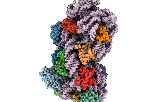 |
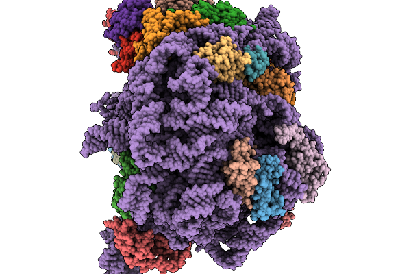
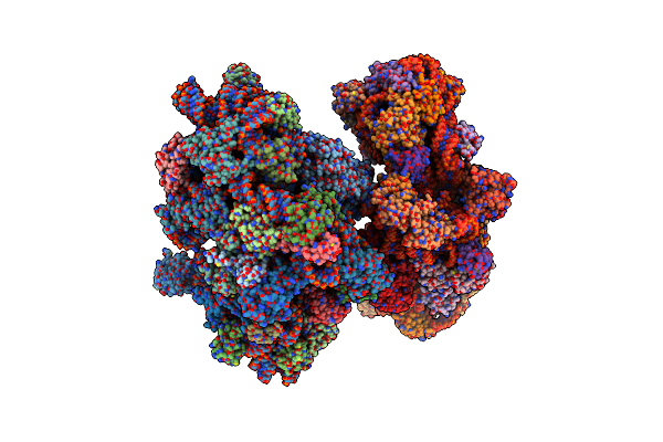
Organism: Escherichia coli
Method: ELECTRON MICROSCOPY Release Date: 2026-02-04 Classification: RIBOSOME |
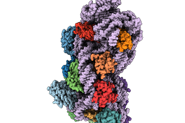 |
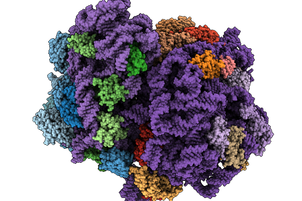
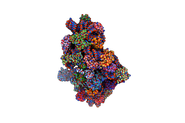
Organism: Escherichia coli
Method: ELECTRON MICROSCOPY Release Date: 2026-02-04 Classification: RIBOSOME |
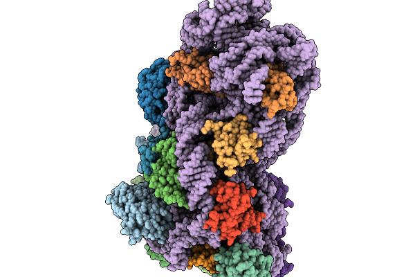 |
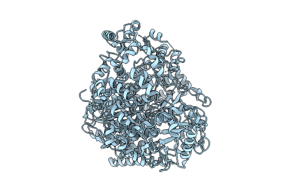
Organism: Escherichia coli
Method: ELECTRON MICROSCOPY Release Date: 2026-02-04 Classification: RIBOSOME |
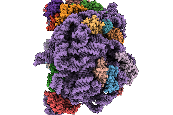 |
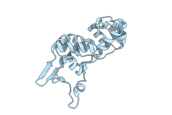
Organism: Escherichia coli
Method: ELECTRON MICROSCOPY Release Date: 2026-02-04 Classification: RIBOSOME |
 |
Organism: Escherichia coli
Method: ELECTRON MICROSCOPY Release Date: 2026-02-04 Classification: RIBOSOME |
 |
Organism: Escherichia coli
Method: ELECTRON MICROSCOPY Release Date: 2026-02-04 Classification: RIBOSOME |
 |
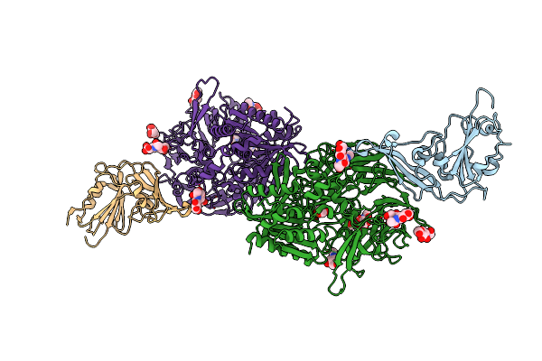
Organism: Rift valley fever virus (strain zh-548 m12)
Method: ELECTRON MICROSCOPY Release Date: 2025-11-12 Classification: VIRAL PROTEIN |
 |
Organism: Geobacillus stearothermophilus
Method: X-RAY DIFFRACTION Resolution:1.49 Å Release Date: 2025-08-27 Classification: RNA BINDING PROTEIN |
 |
Organism: Pyrococcus furiosus
Method: ELECTRON MICROSCOPY Release Date: 2025-01-15 Classification: TRANSLATION |
 |
Organism: Pyrococcus furiosus
Method: ELECTRON MICROSCOPY Release Date: 2025-01-15 Classification: TRANSLATION |
 |
Organism: Pyrococcus furiosus
Method: ELECTRON MICROSCOPY Release Date: 2025-01-15 Classification: TRANSLATION |
 |
Organism: Homo sapiens, Pangolin coronavirus hku4
Method: X-RAY DIFFRACTION Resolution:2.60 Å Release Date: 2024-10-30 Classification: VIRAL PROTEIN Ligands: NAG, EPE |
 |
Organism: Manis javanica, Pangolin coronavirus hku4
Method: X-RAY DIFFRACTION Resolution:2.70 Å Release Date: 2024-10-30 Classification: VIRAL PROTEIN Ligands: NAG, GOL |
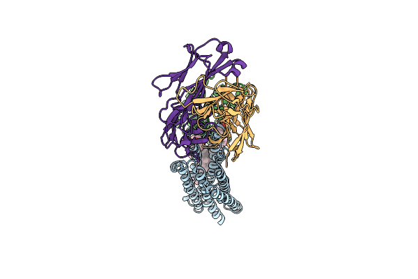 |
Organism: Mus musculus, Homo sapiens
Method: ELECTRON MICROSCOPY Release Date: 2024-09-11 Classification: MEMBRANE PROTEIN/IMMUNE SYSTEM Ligands: CLR |
 |
Organism: Acidaminococcus sp. bv3l6, Synthetic construct
Method: ELECTRON MICROSCOPY Release Date: 2024-07-03 Classification: DNA BINDING PROTEIN/DNA/RNA |
 |
Organism: Acidaminococcus sp. bv3l6, Synthetic construct
Method: ELECTRON MICROSCOPY Release Date: 2024-07-03 Classification: DNA BINDING PROTEIN/DNA/RNA |
 |
Organism: Acidaminococcus sp. bv3l6, Synthetic construct
Method: ELECTRON MICROSCOPY Release Date: 2024-07-03 Classification: DNA BINDING PROTEIN/DNA/RNA |
 |
Organism: Acidaminococcus sp. bv3l6, Synthetic construct
Method: ELECTRON MICROSCOPY Release Date: 2024-07-03 Classification: DNA BINDING PROTEIN/DNA/RNA |
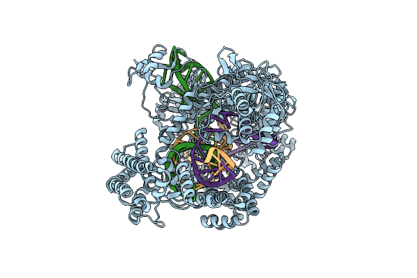 |
Wt Crispr-Cas12A With A 16Bp R-Loop And Nontarget Strand In The Ruvc Active Site.
Organism: Acidaminococcus sp. bv3l6, Synthetic construct
Method: ELECTRON MICROSCOPY Release Date: 2024-07-03 Classification: DNA BINDING PROTEIN/DNA/RNA |
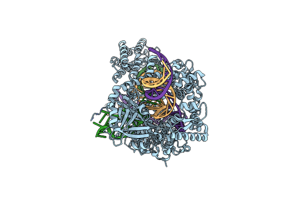 |
Wt Crispr-Cas12A With A 20Bp R-Loop And Nontarget Strand In The Ruvc Active Site.
Organism: Acidaminococcus sp. bv3l6, Synthetic construct
Method: ELECTRON MICROSCOPY Release Date: 2024-07-03 Classification: DNA BINDING PROTEIN/DNA/RNA Ligands: MG |