
Search Count: 28
All
Selected
 |
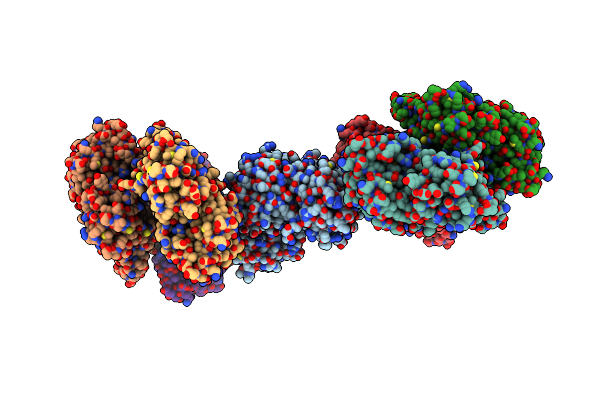
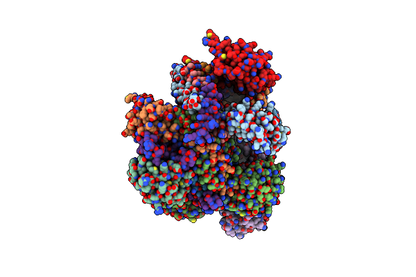
Inner Layer Protein P1 Chains In Transcribing Particles Of Bacteriophage Phi6
Organism: Cystovirus phi6
Method: ELECTRON MICROSCOPY Release Date: 2026-01-21 Classification: VIRAL PROTEIN |
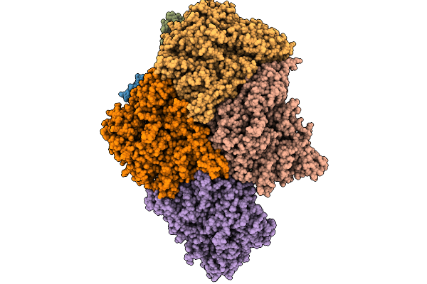 |
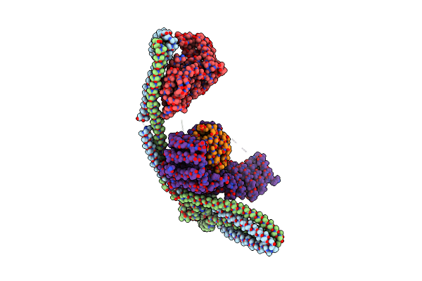
Extended And Wrapped Protein P7 Dimers Of Dimers, The P1 Layer And The Rna-Dependent Rna Polymerase P2 In Transcribing Particles Of Bacteriophage Phi6
Organism: Synthetic construct, Cystovirus phi6
Method: ELECTRON MICROSCOPY Release Date: 2026-01-21 Classification: VIRAL PROTEIN |
 |
Organism: Cystovirus phi6
Method: ELECTRON MICROSCOPY Release Date: 2026-01-14 Classification: VIRUS |
 |
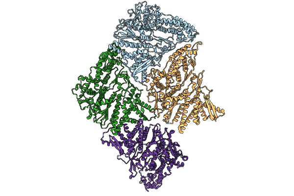
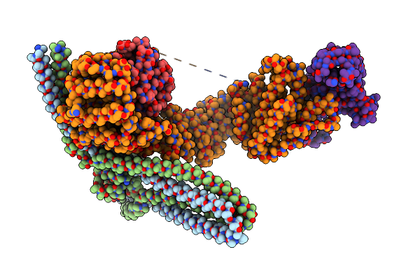
Asymmetric Unit From Transcribing Double-Layered Particle Of Bacteriophage Phi6
Organism: Cystovirus phi6
Method: ELECTRON MICROSCOPY Release Date: 2026-01-14 Classification: VIRUS |
 |
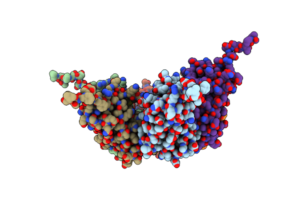
Asymmetric Unit From Transcribing Single-Layered Particle Of Bacteriophage Phi6
Organism: Cystovirus phi6
Method: ELECTRON MICROSCOPY Release Date: 2026-01-14 Classification: VIRUS |
 |
Asymmetric Unit From Transcribing Single-Layered Particle Of Bacteriophage Phi6 In An Over-Expanded State
Organism: Cystovirus phi6
Method: ELECTRON MICROSCOPY Release Date: 2026-01-14 Classification: VIRUS |
 |
Organism: Homo sapiens
Method: ELECTRON MICROSCOPY Release Date: 2025-06-11 Classification: STRUCTURAL PROTEIN |
 |
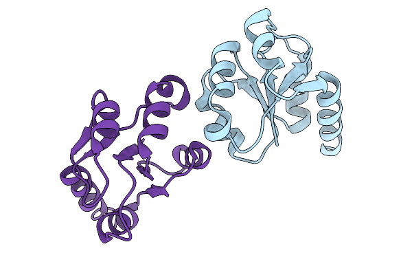
Structure Of The Human Commander Complex Coiled Coils, Dennd10 And Partial Retriever Subcomplex
Organism: Homo sapiens
Method: ELECTRON MICROSCOPY Release Date: 2024-03-20 Classification: UNKNOWN FUNCTION |
 |
Organism: Homo sapiens
Method: ELECTRON MICROSCOPY Release Date: 2024-03-20 Classification: UNKNOWN FUNCTION |
 |
Organism: Homo sapiens
Method: ELECTRON MICROSCOPY Release Date: 2024-03-20 Classification: UNKNOWN FUNCTION |
 |
Organism: Cowpea chlorotic mottle virus
Method: ELECTRON MICROSCOPY Release Date: 2023-07-05 Classification: VIRUS LIKE PARTICLE |
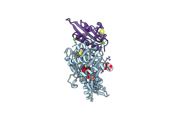 |
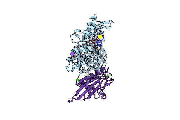
Crystal Structure Of Plasmodium Falciparum Actin I In The Mg-K-Atp/Adp State
Organism: Plasmodium falciparum, Mus musculus
Method: X-RAY DIFFRACTION Resolution:1.24 Å Release Date: 2019-06-26 Classification: CONTRACTILE PROTEIN Ligands: ATP, ADP, MG, K, SCN, BTB, CL, CA |
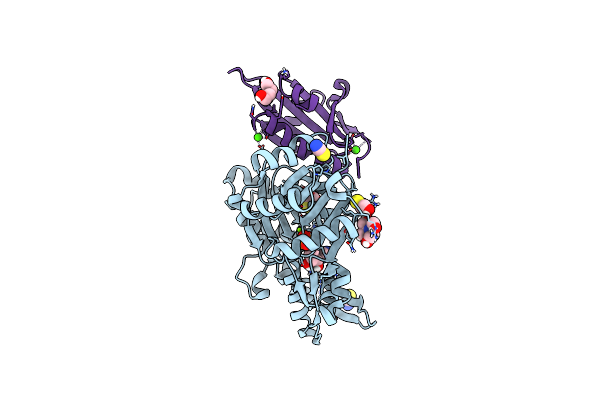 |
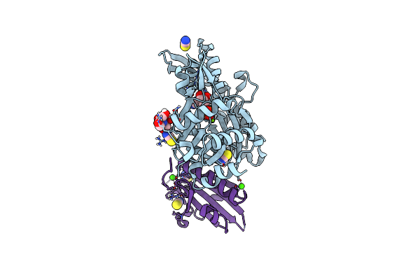
Organism: Plasmodium falciparum, Mus musculus
Method: X-RAY DIFFRACTION Resolution:1.22 Å Release Date: 2019-06-26 Classification: CONTRACTILE PROTEIN Ligands: ADP, MG, CA, SCN, BME, BTB, PEG |
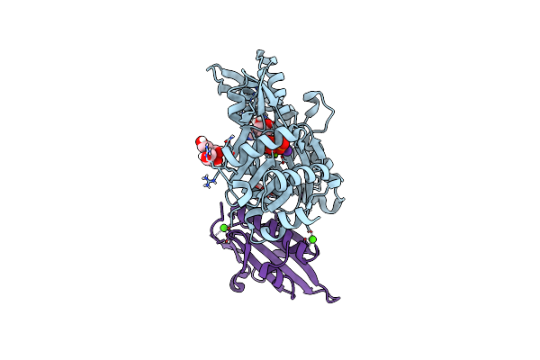 |
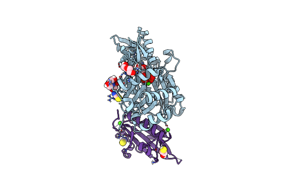
Crystal Structure Of Plasmodium Falciparum Actin I (A272W Mutant) In The Mg-K-Atp/Adp State
Organism: Plasmodium falciparum, Mus musculus
Method: X-RAY DIFFRACTION Resolution:1.50 Å Release Date: 2019-06-26 Classification: CONTRACTILE PROTEIN Ligands: ATP, ADP, MG, K, BTB, PEG, NA, CL, CA |
 |
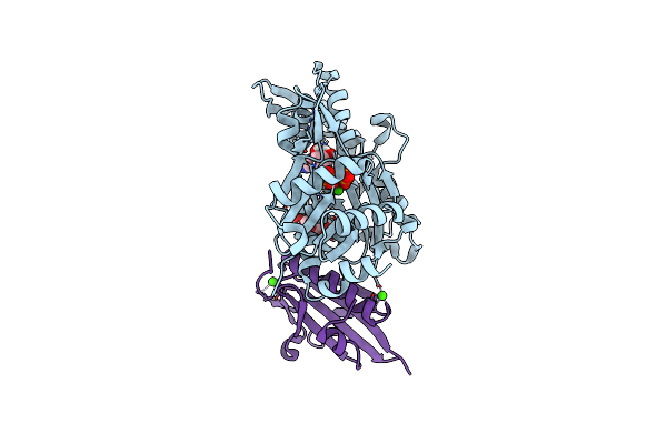
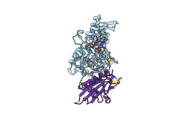
Crystal Structure Of Plasmodium Falciparum Actin I (H74Q) In The Mg-K-Atp State
Organism: Plasmodium falciparum, Mus musculus
Method: X-RAY DIFFRACTION Resolution:2.00 Å Release Date: 2019-06-26 Classification: CONTRACTILE PROTEIN Ligands: ATP, MG, K, BME, CA, SCN |
 |
Crystal Structure Of Plasmodium Falciparum Actin I (F54Y Mutant) In The Ca-Atp State
Organism: Plasmodium falciparum, Mus musculus
Method: X-RAY DIFFRACTION Resolution:1.40 Å Release Date: 2019-06-26 Classification: CONTRACTILE PROTEIN Ligands: ATP, CA, CL, PEG |
 |
Crystal Structure Of Plasmodium Falciparum Actin I (F54Y Mutant) In The Mg-K-Adp-Alfn State
Organism: Plasmodium falciparum, Mus musculus
Method: X-RAY DIFFRACTION Resolution:1.90 Å Release Date: 2019-06-26 Classification: CONTRACTILE PROTEIN Ligands: ADP, AF3, MG, K, SCN, CA |
 |
Crystal Structure Of Plasmodium Falciparum Actin I (F54Y Mutant) In The Mg-Adp State
Organism: Plasmodium falciparum, Mus musculus
Method: X-RAY DIFFRACTION Resolution:1.50 Å Release Date: 2019-06-26 Classification: CONTRACTILE PROTEIN Ligands: ADP, MG, SCN, BTB, K, CA |
 |
Crystal Structure Of Plasmodium Falciparum Actin I (G115A Mutant) In The Ca-Atp State
Organism: Plasmodium falciparum, Mus musculus
Method: X-RAY DIFFRACTION Resolution:1.83 Å Release Date: 2019-06-26 Classification: CONTRACTILE PROTEIN Ligands: ATP, CA, SCN, BTB, PEG, CL, BME |
 |
Crystal Structure Of Plasmodium Falciparum Actin I (G115A Mutant) In The Mg-K-Atp/Adp State
Organism: Plasmodium falciparum, Mus musculus
Method: X-RAY DIFFRACTION Resolution:1.83 Å Release Date: 2019-06-26 Classification: CONTRACTILE PROTEIN Ligands: ATP, ADP, MG, SCN, CA |