

Search Count: 39
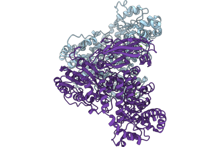 |
Crystal Structure Of Beta-Glucosidase From Bacteroides Ovatus
Organism: Bacteroides ovatus atcc 8483
Method: X-RAY DIFFRACTION Resolution:1.40 Å Release Date: 2026-05-20 Classification: HYDROLASE Ligands: MG |
Organism: Bacteroides ovatus atcc 8483
Method: X-RAY DIFFRACTION
Release Date: 2026-05-20
Ligands: MG
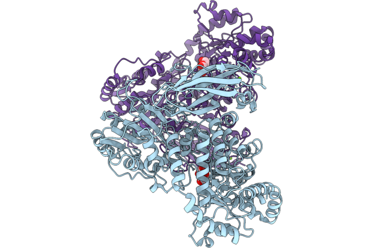 |
Crystal Structure Of Beta-Glucosidase From Bacteroides Ovatus Bound To Beta-D-Glucose
Organism: Bacteroides ovatus atcc 8483
Method: X-RAY DIFFRACTION Resolution:1.95 Å Release Date: 2026-05-20 Classification: HYDROLASE Ligands: BGC, MG |
Organism: Bacteroides ovatus atcc 8483
Method: X-RAY DIFFRACTION
Release Date: 2026-05-20
Ligands: BGC, MG
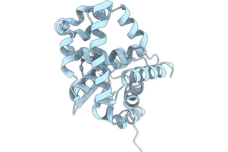 |
Crystal Structure Of Zinc-Binding Metallopeptidase (Uniprot: A0Aan3A8D2) From Bacteroides Ovatus
Organism: Bacteroides ovatus atcc 8483
Method: X-RAY DIFFRACTION Resolution:2.10 Å Release Date: 2026-04-29 Classification: HYDROLASE Ligands: ZN |
Organism: Bacteroides ovatus atcc 8483
Method: X-RAY DIFFRACTION
Release Date: 2026-04-29
Ligands: ZN
 |
Crystal Structure Of Procerain-B From Calotropis Gigantea
Organism: Calotropis
Method: X-RAY DIFFRACTION Resolution:1.50 Å Release Date: 2025-04-23 Classification: HYDROLASE |
Organism: Calotropis
Method: X-RAY DIFFRACTION
Release Date: 2025-04-23
 |
Crystal Structure Of Procerain-B From Calotropis Gigantea With Glycerol
Organism: Calotropis
Method: X-RAY DIFFRACTION Resolution:1.90 Å Release Date: 2025-04-23 Classification: HYDROLASE |
Organism: Calotropis
Method: X-RAY DIFFRACTION
Release Date: 2025-04-23
 |
Crystal Structure Of Calotropain Fi From Calotropis Gigantea
Organism: Calotropis gigantea
Method: X-RAY DIFFRACTION Resolution:1.25 Å Release Date: 2024-05-22 Classification: HYDROLASE Ligands: E64 |
Organism: Calotropis gigantea
Method: X-RAY DIFFRACTION
Release Date: 2024-05-22
Ligands: E64
 |
Crystal Structure Of Calotropain Fi From Calotropis Gigantea (Ph 6.0)
Organism: Calotropis gigantea
Method: X-RAY DIFFRACTION Resolution:1.40 Å Release Date: 2024-05-22 Classification: HYDROLASE Ligands: GOL, E64 |
Organism: Calotropis gigantea
Method: X-RAY DIFFRACTION
Release Date: 2024-05-22
Ligands: GOL, E64
 |
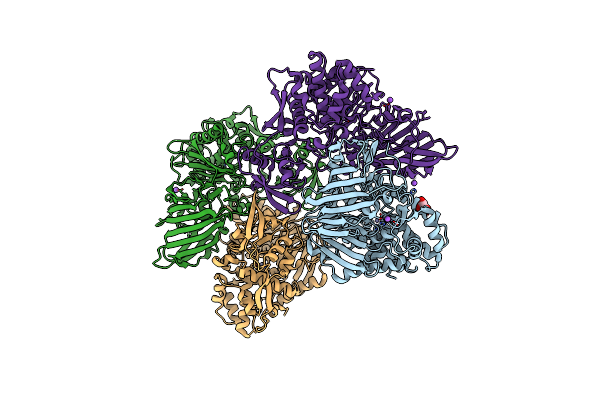
Crystal Structure Of Procerain-B From Calotropis Gigantea
Organism: Calotropis
Method: X-RAY DIFFRACTION Resolution:1.25 Å Release Date: 2024-05-22 Classification: HYDROLASE |
Organism: Calotropis
Method: X-RAY DIFFRACTION
Release Date: 2024-05-22
 |
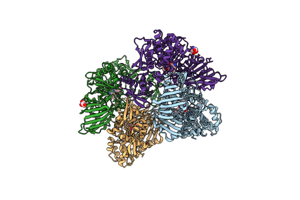
Crystal Structure Of M61 Peptidase (Apo-Form) From Xanthomonas Campestris
Organism: Xanthomonas campestris pv. campestris b100
Method: X-RAY DIFFRACTION Resolution:2.10 Å Release Date: 2024-05-01 Classification: HYDROLASE Ligands: GOL, ZN, NA |
Organism: Xanthomonas campestris pv. campestris b100
Method: X-RAY DIFFRACTION
Release Date: 2024-05-01
Ligands: GOL, ZN, NA
 |
Crystal Structure Of M61 Peptidase (Bestatin-Bound) From Xanthomonas Campestris
Organism: Xanthomonas campestris pv. campestris b100
Method: X-RAY DIFFRACTION Resolution:1.90 Å Release Date: 2024-05-01 Classification: HYDROLASE Ligands: ZN, BES, GOL, TRS |
Organism: Xanthomonas campestris pv. campestris b100
Method: X-RAY DIFFRACTION
Release Date: 2024-05-01
Ligands: ZN, BES, GOL, TRS
 |
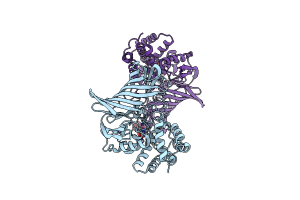
Crystal Structure Of Two Domain M1 Zinc Metallopeptidase E323A Mutant Bound To L-Methionine Amino Acid
Organism: Deinococcus radiodurans (strain atcc 13939 / dsm 20539 / jcm 16871 / lmg 4051 / nbrc 15346 / ncimb 9279 / r1 / vkm b-1422)
Method: X-RAY DIFFRACTION Resolution:2.19 Å Release Date: 2020-06-24 Classification: HYDROLASE Ligands: ZN, NA, MET |
 |
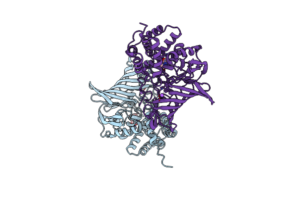
Crystal Structure Of Two Domain M1 Zinc Metallopeptidase E323A Mutant Bound To L-Tryptophan Amino Acid
Organism: Deinococcus radiodurans (strain atcc 13939 / dsm 20539 / jcm 16871 / lmg 4051 / nbrc 15346 / ncimb 9279 / r1 / vkm b-1422)
Method: X-RAY DIFFRACTION Resolution:2.35 Å Release Date: 2020-01-22 Classification: HYDROLASE Ligands: TRP, ZN |
 |
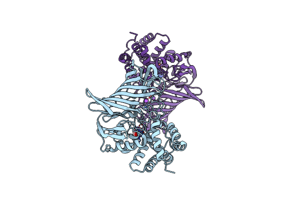
Crystal Structure Of Two Domain M1 Zinc Metallopeptidase E323 Mutant Bound To L-Leucine Amino Acid
Organism: Deinococcus radiodurans (strain atcc 13939 / dsm 20539 / jcm 16871 / lmg 4051 / nbrc 15346 / ncimb 9279 / r1 / vkm b-1422)
Method: X-RAY DIFFRACTION Resolution:2.25 Å Release Date: 2020-01-22 Classification: HYDROLASE Ligands: ZN, LEU, NA |
 |
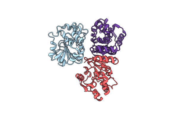
Crystal Structure Of Two Domain M1 Zinc Metallopeptidase E323A Mutant Bound To L-Arginine
Organism: Deinococcus radiodurans (strain atcc 13939 / dsm 20539 / jcm 16871 / lmg 4051 / nbrc 15346 / ncimb 9279 / r1 / vkm b-1422)
Method: X-RAY DIFFRACTION Resolution:2.10 Å Release Date: 2020-01-22 Classification: HYDROLASE Ligands: ZN, ARG, NA |
 |
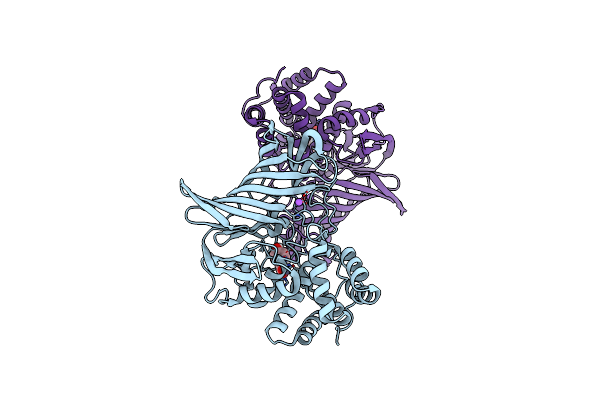
Crystal Structure Of Peptidase E From Deinococcus Radiodurans In P6422 Space Group
Organism: Deinococcus radiodurans
Method: X-RAY DIFFRACTION Resolution:2.70 Å Release Date: 2019-11-20 Classification: HYDROLASE |
Organism: Deinococcus radiodurans
Method: X-RAY DIFFRACTION
Release Date: 2019-11-20
 |
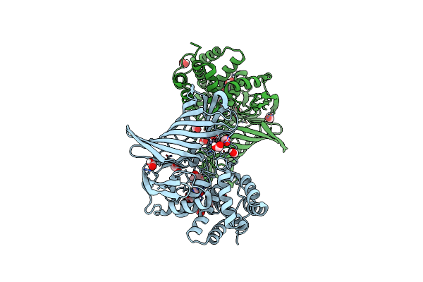
Crystal Structure Of M1 Zinc Metallopeptidase E323A Mutant From Deinococcus Radiodurans
Organism: Deinococcus radiodurans (strain atcc 13939 / dsm 20539 / jcm 16871 / lmg 4051 / nbrc 15346 / ncimb 9279 / r1 / vkm b-1422)
Method: X-RAY DIFFRACTION Resolution:1.83 Å Release Date: 2019-09-25 Classification: HYDROLASE Ligands: ZN, TYR, NA |
 |
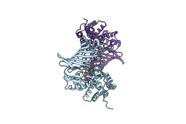
Crystal Structure Of M1 Zinc Metallopeptidase E323A Mutant Bound To Tyr-Ser-Ala Substrate From Deinococcus Radiodurans
Organism: Deinococcus radiodurans (strain atcc 13939 / dsm 20539 / jcm 16871 / lmg 4051 / nbrc 15346 / ncimb 9279 / r1 / vkm b-1422), Deinococcus
Method: X-RAY DIFFRACTION Resolution:1.90 Å Release Date: 2019-09-25 Classification: HYDROLASE Ligands: ZN, FMT |
 |
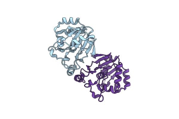
Crystal Structure Of M1 Zinc Metallopeptidase From Deinococcus Radiodurans
Organism: Deinococcus radiodurans (strain atcc 13939 / dsm 20539 / jcm 16871 / lmg 4051 / nbrc 15346 / ncimb 9279 / r1 / vkm b-1422)
Method: X-RAY DIFFRACTION Resolution:2.05 Å Release Date: 2019-07-17 Classification: HYDROLASE Ligands: ZN, TYR, NA |
 |
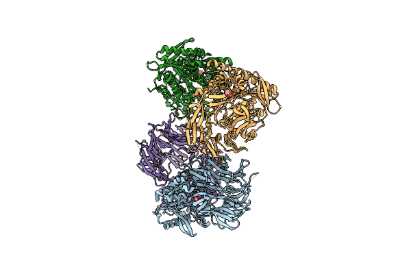
Crystal Structure Of Peptidase E From Deinococcus Radiodurans R1
Organism: Deinococcus radiodurans r1
Method: X-RAY DIFFRACTION Resolution:2.00 Å Release Date: 2019-06-26 Classification: HYDROLASE |
Organism: Deinococcus radiodurans r1
Method: X-RAY DIFFRACTION
Release Date: 2019-06-26
 |
Crystal Structure Of S9 Peptidase (Inactive Form) From Deinococcus Radiodurans R1
Organism: Deinococcus radiodurans (strain atcc 13939 / dsm 20539 / jcm 16871 / lmg 4051 / nbrc 15346 / ncimb 9279 / r1 / vkm b-1422)
Method: X-RAY DIFFRACTION Resolution:2.30 Å Release Date: 2018-11-14 Classification: HYDROLASE Ligands: ACT |