

Search Count: 35
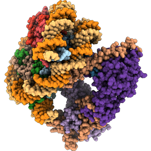 |
Drosophila Msl Hat Nucleosome Complex H4K16Ac Post-Catalytic State
Organism: Homo sapiens, Drosophila melanogaster, Synthetic construct
Method: ELECTRON MICROSCOPY Release Date: 2026-10-14 Classification: NUCLEAR PROTEIN Ligands: ZN, COA |
Organism: Homo sapiens, Drosophila melanogaster, Synthetic construct
Method: ELECTRON MICROSCOPY
Release Date: 2026-10-14
Ligands: ZN, COA
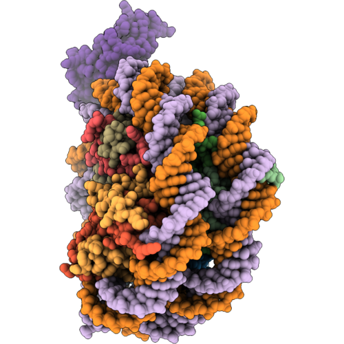 |
Drosophila Msl Hat Nucleosome Complex H3K36Me3 Reader State
Organism: Homo sapiens, Drosophila melanogaster, Synthetic construct
Method: ELECTRON MICROSCOPY Release Date: 2026-10-14 Classification: NUCLEAR PROTEIN |
Organism: Homo sapiens, Drosophila melanogaster, Synthetic construct
Method: ELECTRON MICROSCOPY
Release Date: 2026-10-14
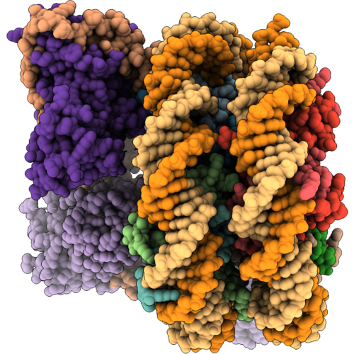 |
Drosophila Msl Hat Nucleosome Complex H4K16Ac Postcatalytic H3K36Me3 Reader State
Organism: Homo sapiens, Drosophila melanogaster, Synthetic construct
Method: ELECTRON MICROSCOPY Release Date: 2026-10-14 Classification: NUCLEAR PROTEIN Ligands: ZN, COA |
Organism: Homo sapiens, Drosophila melanogaster, Synthetic construct
Method: ELECTRON MICROSCOPY
Release Date: 2026-10-14
Ligands: ZN, COA
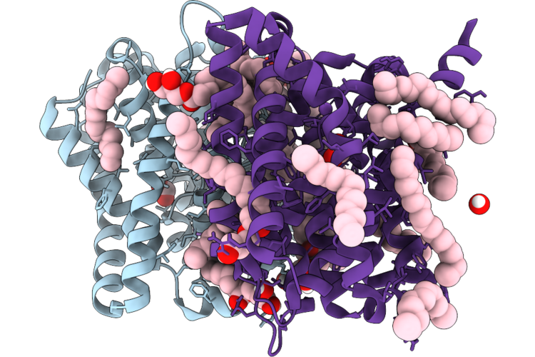 |
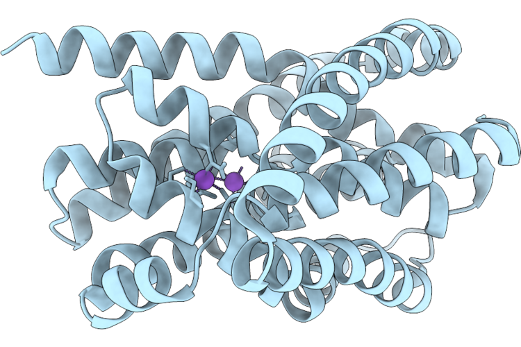
Structure Of Asbt Homologue From Leptospira Biflexa In Inward-Facing Form (Crystal Form 1)
Organism: Leptospira biflexa
Method: X-RAY DIFFRACTION Resolution:2.21 Å Release Date: 2026-07-22 Classification: MEMBRANE PROTEIN |
Organism: Leptospira biflexa
Method: X-RAY DIFFRACTION
Release Date: 2026-07-22
 |
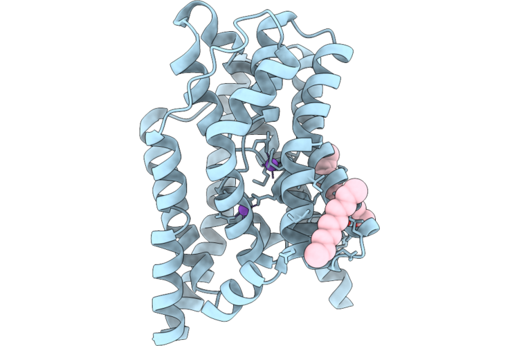
Structure Of Asbt Homologue From Leptospira Biflexa In Inward-Facing Form (Crystal Form 2)
Organism: Leptospira biflexa
Method: X-RAY DIFFRACTION Resolution:2.92 Å Release Date: 2026-07-22 Classification: MEMBRANE PROTEIN |
Organism: Leptospira biflexa
Method: X-RAY DIFFRACTION
Release Date: 2026-07-22
 |
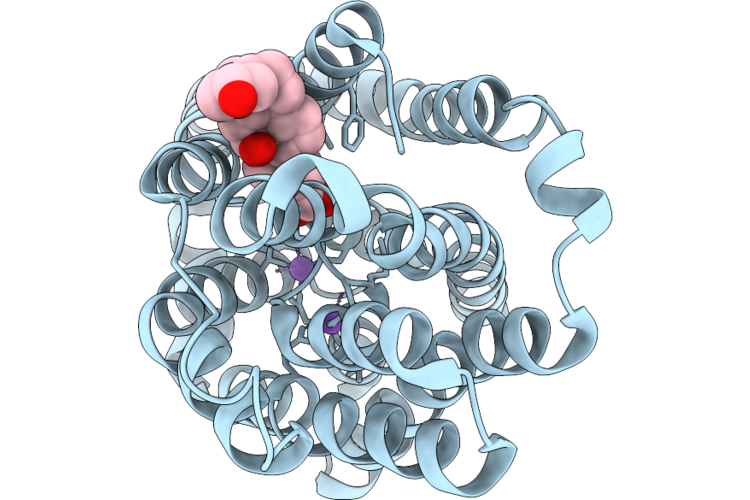
Structure Of Mutated Asbt Homologue From Leptospira Biflexa In Outward-Facing Form (No Bile Acid)
Organism: Leptospira biflexa
Method: X-RAY DIFFRACTION Resolution:2.59 Å Release Date: 2026-07-22 Classification: MEMBRANE PROTEIN |
Organism: Leptospira biflexa
Method: X-RAY DIFFRACTION
Release Date: 2026-07-22
 |
Structure Of Mutated Asbt Homologue From Leptospira Biflexa In Outward-Facing Form With Deoxycholate
Organism: Leptospira biflexa
Method: X-RAY DIFFRACTION Resolution:2.52 Å Release Date: 2026-07-22 Classification: MEMBRANE PROTEIN |
Organism: Leptospira biflexa
Method: X-RAY DIFFRACTION
Release Date: 2026-07-22
 |
Cryo-Em Structure Of Mle In Complex With Sl7Uuc Rna And Adp
Organism: Drosophila melanogaster
Method: ELECTRON MICROSCOPY Release Date: 2023-11-22 Classification: RNA BINDING PROTEIN Ligands: ADP |
Organism: Drosophila melanogaster
Method: ELECTRON MICROSCOPY
Release Date: 2023-11-22
Ligands: ADP
 |
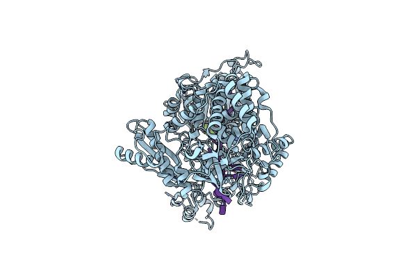
Cryo-Em Structure Of Mle In Complex With Uuc Rna And Adp
Organism: Drosophila melanogaster
Method: ELECTRON MICROSCOPY Release Date: 2023-11-01 Classification: RNA BINDING PROTEIN Ligands: ADP, MG |
Organism: Drosophila melanogaster
Method: ELECTRON MICROSCOPY
Release Date: 2023-11-01
Ligands: ADP, MG
 |
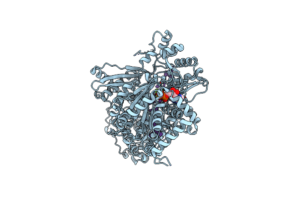
Cryo-Em Structure Of Mle In Complex With Adp:Alf4 And U10 Rna
Organism: Drosophila melanogaster
Method: ELECTRON MICROSCOPY Release Date: 2023-10-18 Classification: RNA BINDING PROTEIN Ligands: ADP, ALF |
Organism: Drosophila melanogaster
Method: ELECTRON MICROSCOPY
Release Date: 2023-10-18
Ligands: ADP, ALF
 |
Cryo-Em Structure Of Mle In Complex With Adp:Alf4 And Uuc Rna
Organism: Drosophila melanogaster
Method: ELECTRON MICROSCOPY Release Date: 2023-10-18 Classification: RNA BINDING PROTEIN Ligands: ADP, MG, ALF |
Organism: Drosophila melanogaster
Method: ELECTRON MICROSCOPY
Release Date: 2023-10-18
Ligands: ADP, MG, ALF
 |
Cryo-Em Structure Of Mle In Complex With Adp:Alf4
Organism: Drosophila melanogaster
Method: ELECTRON MICROSCOPY Release Date: 2023-10-18 Classification: RNA BINDING PROTEIN Ligands: ADP, ALF, MG |
Organism: Drosophila melanogaster
Method: ELECTRON MICROSCOPY
Release Date: 2023-10-18
Ligands: ADP, ALF, MG
 |
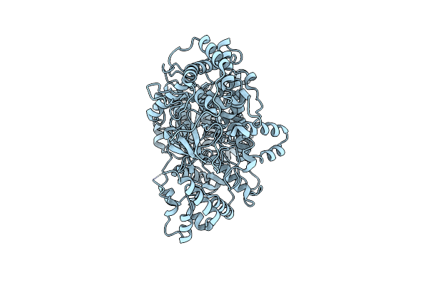
Cryo-Em Structure Of Mle In Complex With Adp:Alf4 And Sl7Moduuc Rna
Organism: Drosophila melanogaster
Method: ELECTRON MICROSCOPY Release Date: 2023-10-18 Classification: RNA BINDING PROTEIN Ligands: ADP, ALF |
Organism: Drosophila melanogaster
Method: ELECTRON MICROSCOPY
Release Date: 2023-10-18
Ligands: ADP, ALF
 |
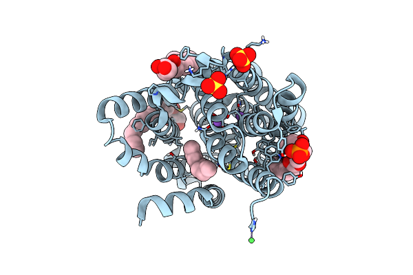
Cryo-Em Structure Of Mle
Organism: Drosophila melanogaster
Method: ELECTRON MICROSCOPY Release Date: 2023-10-18 Classification: RNA BINDING PROTEIN |
Organism: Drosophila melanogaster
Method: ELECTRON MICROSCOPY
Release Date: 2023-10-18
 |
Crystal Structure Of Asbtnm In Lipidic Cubic Phase Without Substrate Bound
Organism: Neisseria meningitidis mc58
Method: X-RAY DIFFRACTION Resolution:2.10 Å Release Date: 2023-07-12 Classification: TRANSPORT PROTEIN |
Organism: Neisseria meningitidis mc58
Method: X-RAY DIFFRACTION
Release Date: 2023-07-12
 |
Crystal Structure Of Asbtnm In Complex With Pantoate
Organism: Neisseria meningitidis mc58
Method: X-RAY DIFFRACTION Resolution:2.30 Å Release Date: 2023-07-12 Classification: TRANSPORT PROTEIN |
Organism: Neisseria meningitidis mc58
Method: X-RAY DIFFRACTION
Release Date: 2023-07-12
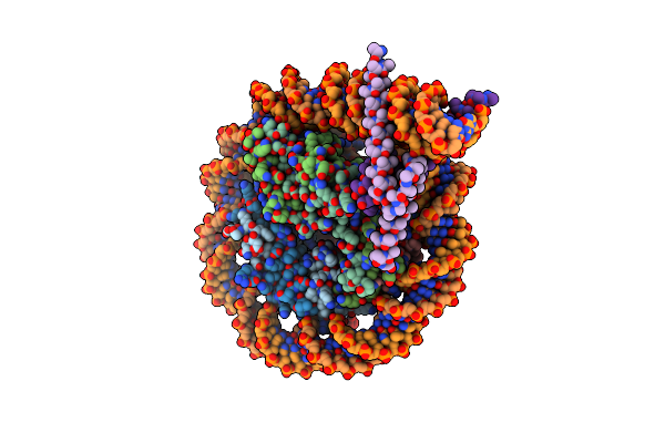 |
Cryo-Em Structure Of Clock-Bmal1 Bound To A Nucleosomal E-Box At Position Shl-6.2 (Dna Conformation 1)
Organism: Homo sapiens, Mus musculus, Synthetic construct
Method: ELECTRON MICROSCOPY Release Date: 2023-05-24 Classification: GENE REGULATION |
Organism: Homo sapiens, Mus musculus, Synthetic construct
Method: ELECTRON MICROSCOPY
Release Date: 2023-05-24
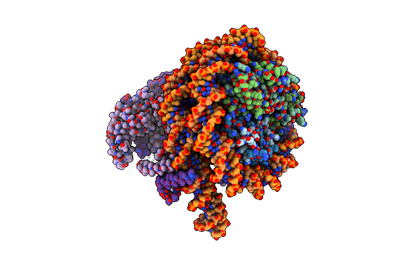 |
Cryo-Em Structure Of Clock-Bmal1 Bound To A Nucleosomal E-Box At Position Shl+5.8 (Composite Map)
Organism: Homo sapiens, Mus musculus, Synthetic construct
Method: ELECTRON MICROSCOPY Release Date: 2023-05-24 Classification: GENE REGULATION |
Organism: Homo sapiens, Mus musculus, Synthetic construct
Method: ELECTRON MICROSCOPY
Release Date: 2023-05-24
 |
Cryo-Em Structure Of Clock-Bmal1 Bound To The Native Por Enhancer Nucleosome (Map 2, Additional 3D Classification And Flexible Refinement)
Organism: Homo sapiens, Mus musculus
Method: ELECTRON MICROSCOPY Release Date: 2023-05-24 Classification: GENE REGULATION |
Organism: Homo sapiens, Mus musculus
Method: ELECTRON MICROSCOPY
Release Date: 2023-05-24
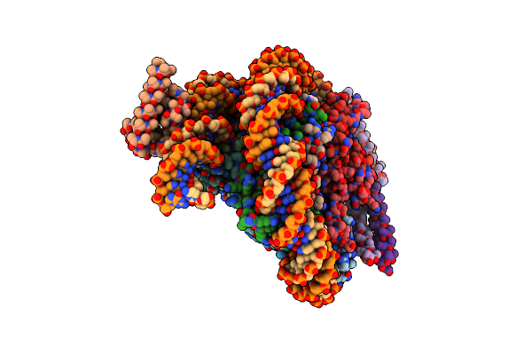 |
Oct4 And Myc-Max Co-Bound To A Nucleosome
Organism: Homo sapiens, Aequorea victoria, Synthetic construct
Method: ELECTRON MICROSCOPY Release Date: 2023-05-24 Classification: TRANSCRIPTION Ligands: PTD |
Organism: Homo sapiens, Aequorea victoria, Synthetic construct
Method: ELECTRON MICROSCOPY
Release Date: 2023-05-24
Ligands: PTD