
Search Count: 1,616
 |
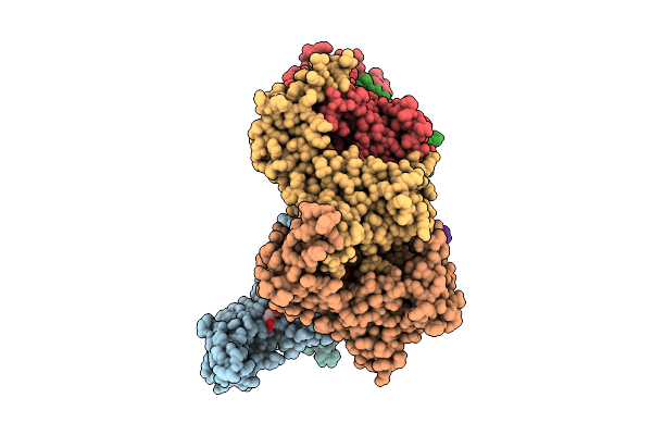
Apo Crystal Structure Of A Computationally Designed Protein (Trp)
Organism: Escherichia coli bl21(de3)
Method: X-RAY DIFFRACTION Resolution:2.10 Å Release Date: 2026-06-03 Classification: DE NOVO PROTEIN |
Organism: Escherichia coli bl21(de3)
Method: X-RAY DIFFRACTION
Release Date: 2026-06-03
 |
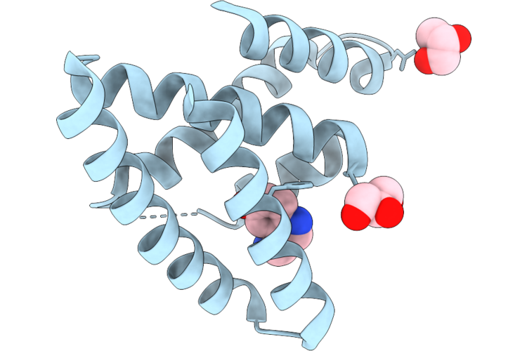
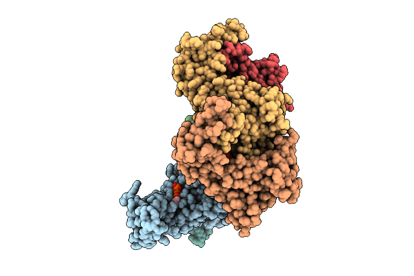
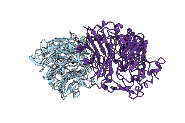
Crystal Structure Of De Novo Designed Serotonin Binder Srob2_30
Organism: Escherichia coli bl21(de3)
Method: X-RAY DIFFRACTION Resolution:2.58 Å Release Date: 2026-05-27 Classification: DE NOVO PROTEIN Ligands: EPE, SRO |
Organism: Escherichia coli bl21(de3)
Method: X-RAY DIFFRACTION
Release Date: 2026-05-27
Ligands: EPE, SRO
 |
Crystal Structure Of De Novo Designed Serotonin Binder Srob2_26_L7F
Organism: Escherichia coli bl21(de3)
Method: X-RAY DIFFRACTION Resolution:2.00 Å Release Date: 2026-05-27 Classification: DE NOVO PROTEIN Ligands: SRO, GOL |
Organism: Escherichia coli bl21(de3)
Method: X-RAY DIFFRACTION
Release Date: 2026-05-27
Ligands: SRO, GOL
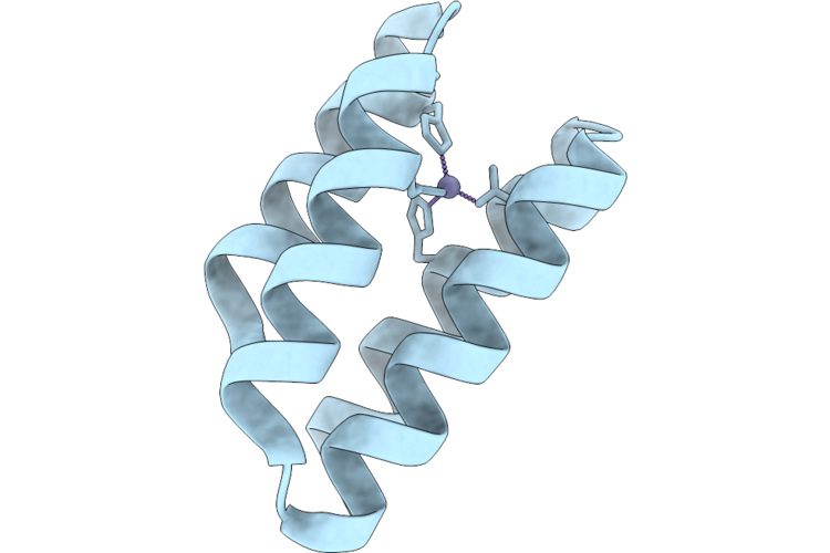 |
Crustal Structure Of De Novo Designed Zinc Binding Protein Zk2
Organism: Escherichia coli bl21(de3)
Method: X-RAY DIFFRACTION Resolution:1.30 Å Release Date: 2026-05-27 Classification: DE NOVO PROTEIN Ligands: ZN |
Organism: Escherichia coli bl21(de3)
Method: X-RAY DIFFRACTION
Release Date: 2026-05-27
Ligands: ZN
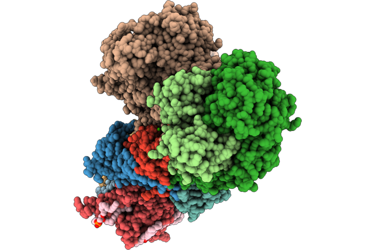 |
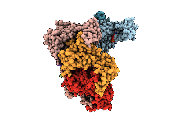
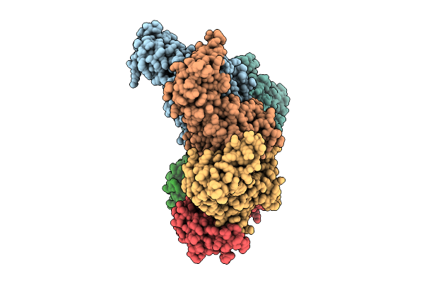
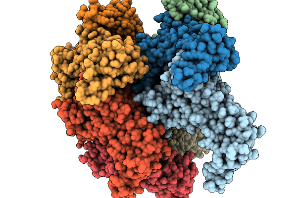
E. Coli Complex I Wt Purified In Lmng
Organism: Escherichia coli bw25113, Escherichia coli bl21(de3)
Method: ELECTRON MICROSCOPY Release Date: 2026-05-06 Classification: PROTON TRANSPORT Ligands: FES, CA, FMN, SF4, 3PE, LFA, CDL, TRD, UQ8 |
Organism: Escherichia coli bw25113, Escherichia coli bl21(de3)
Method: ELECTRON MICROSCOPY
Release Date: 2026-05-06
Ligands: FES, CA, FMN, SF4, 3PE, LFA, CDL, TRD, UQ8
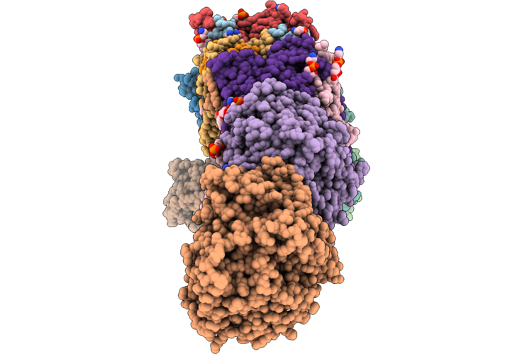 |
E. Coli Complex I D79N Nuoa Mutant Purified In Lmng
Organism: Escherichia coli bl21(de3), Escherichia coli bw25113
Method: ELECTRON MICROSCOPY Release Date: 2026-05-06 Classification: PROTON TRANSPORT Ligands: FES, SF4, FMN, CA, 3PE, 7PH, UQ8 |
Organism: Escherichia coli bl21(de3), Escherichia coli bw25113
Method: ELECTRON MICROSCOPY
Release Date: 2026-05-06
Ligands: FES, SF4, FMN, CA, 3PE, 7PH, UQ8
 |
Dimer Of Atpase Brxc Containing A Walker B Mutation And Bound To Atp From The Acinetobacter Brex System
Organism: Acinetobacter
Method: ELECTRON MICROSCOPY Release Date: 2026-04-08 Classification: ANTIMICROBIAL PROTEIN Ligands: ATP, MG |
Organism: Acinetobacter
Method: ELECTRON MICROSCOPY
Release Date: 2026-04-08
Ligands: ATP, MG
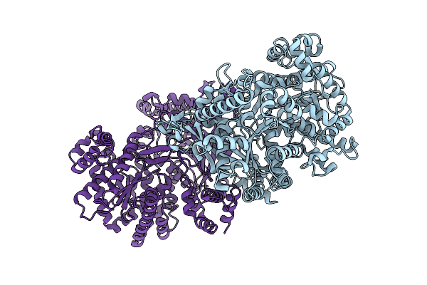 |
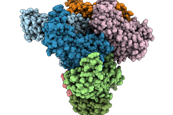
Glycogen Phosphorylase Tetramer From E. Coli
Organism: Escherichia coli bl21(de3)
Method: ELECTRON MICROSCOPY Release Date: 2026-04-01 Classification: STRUCTURAL PROTEIN |
Organism: Escherichia coli bl21(de3)
Method: ELECTRON MICROSCOPY
Release Date: 2026-04-01
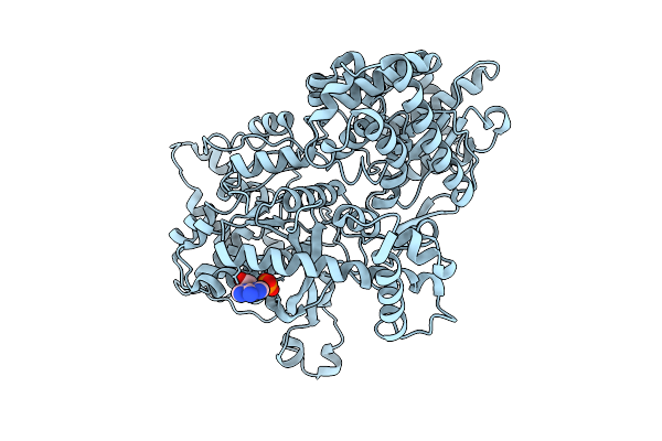 |
Glycogen Phosphorylase Dimer From E. Coli In Complex With Amp.
Organism: Escherichia coli bl21(de3)
Method: ELECTRON MICROSCOPY Release Date: 2026-04-01 Classification: STRUCTURAL PROTEIN Ligands: AMP |
Organism: Escherichia coli bl21(de3)
Method: ELECTRON MICROSCOPY
Release Date: 2026-04-01
Ligands: AMP
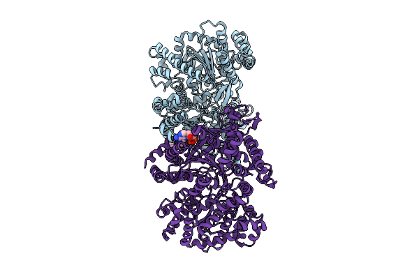 |
Glycogen Phosphorylase Tetramer From E. Coli In Complex With Amp
Organism: Escherichia coli bl21(de3)
Method: ELECTRON MICROSCOPY Release Date: 2026-04-01 Classification: STRUCTURAL PROTEIN Ligands: AMP |
Organism: Escherichia coli bl21(de3)
Method: ELECTRON MICROSCOPY
Release Date: 2026-04-01
Ligands: AMP
 |
Heptamer Msp1 From S.Cerevisiae (With A Catalytic Dead Mutation) In Complex With An Unknown Peptide Substrate
Organism: Saccharomyces cerevisiae s288c, Escherichia coli bl21(de3)
Method: ELECTRON MICROSCOPY Release Date: 2026-03-18 Classification: MEMBRANE PROTEIN Ligands: ATP, MG |
Organism: Saccharomyces cerevisiae s288c, Escherichia coli bl21(de3)
Method: ELECTRON MICROSCOPY
Release Date: 2026-03-18
Ligands: ATP, MG
 |
Hexamer Msp1 From S.Cerevisiae (With A Catalytic Dead Mutation) In Complex With An Unknown Peptide Substrate
Organism: Saccharomyces cerevisiae s288c, Escherichia coli bl21(de3)
Method: ELECTRON MICROSCOPY Release Date: 2026-03-11 Classification: MEMBRANE PROTEIN Ligands: ATP, MG |
Organism: Saccharomyces cerevisiae s288c, Escherichia coli bl21(de3)
Method: ELECTRON MICROSCOPY
Release Date: 2026-03-11
Ligands: ATP, MG
 |
Octamer Msp1 From S.Cerevisiae (With A Catalytic Dead Mutation) In Complex With An Unknown Peptide Substrate
Organism: Saccharomyces cerevisiae s288c, Escherichia coli bl21(de3)
Method: ELECTRON MICROSCOPY Release Date: 2026-03-04 Classification: MEMBRANE PROTEIN Ligands: ATP, MG |
Organism: Saccharomyces cerevisiae s288c, Escherichia coli bl21(de3)
Method: ELECTRON MICROSCOPY
Release Date: 2026-03-04
Ligands: ATP, MG
 |
Nonamer Msp1 From S.Cerevisiae (With A Catalytic Dead Mutation) In Complex With An Unknown Peptide Substrate
Organism: Saccharomyces cerevisiae s288c, Escherichia coli bl21(de3)
Method: ELECTRON MICROSCOPY Release Date: 2026-03-04 Classification: MEMBRANE PROTEIN Ligands: ATP, MG |
Organism: Saccharomyces cerevisiae s288c, Escherichia coli bl21(de3)
Method: ELECTRON MICROSCOPY
Release Date: 2026-03-04
Ligands: ATP, MG
 |
Decamer Msp1 From S.Cerevisiae(With A Catalytic Dead Mutation) In Complex With An Unknown Peptide Substrate
Organism: Saccharomyces cerevisiae s288c, Escherichia coli bl21(de3)
Method: ELECTRON MICROSCOPY Release Date: 2026-03-04 Classification: MEMBRANE PROTEIN Ligands: ATP, MG |
Organism: Saccharomyces cerevisiae s288c, Escherichia coli bl21(de3)
Method: ELECTRON MICROSCOPY
Release Date: 2026-03-04
Ligands: ATP, MG
 |
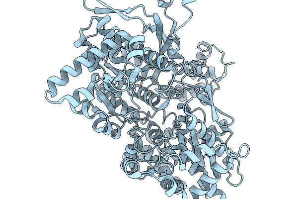
Pqq-Dependent Alcohol Dehydrogenase Detoxifying Don
Organism: Devosia
Method: X-RAY DIFFRACTION Resolution:1.92 Å Release Date: 2026-02-18 Classification: OXIDOREDUCTASE |
Organism: Devosia
Method: X-RAY DIFFRACTION
Release Date: 2026-02-18
 |
Glycogen Phosphorylase Dimer From E. Coli
Organism: Escherichia coli bl21(de3)
Method: ELECTRON MICROSCOPY Release Date: 2026-02-18 Classification: STRUCTURAL PROTEIN |
Organism: Escherichia coli bl21(de3)
Method: ELECTRON MICROSCOPY
Release Date: 2026-02-18
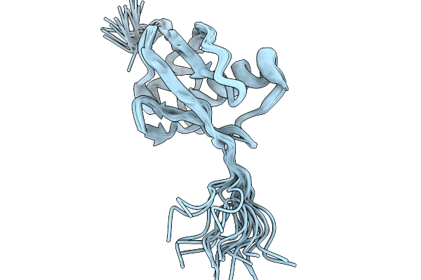 |
Nmr Structure Of Proteinmpnn-Desighed Ubiquitin Variant R4 At Ph 3 With 8 M Urea
Organism: Escherichia coli bl21(de3)
Method: SOLUTION NMR Release Date: 2026-02-11 Classification: DE NOVO PROTEIN |
Organism: Escherichia coli bl21(de3)
Method: SOLUTION NMR
Release Date: 2026-02-11